
1
Assess
Document the mole and evaluate the cheek both at rest and during facial movement.
Mole Removal • Transformation Story
A four-month transformation shaped by facial movement, scar-conscious planning and a carefully executed multilayer closure.
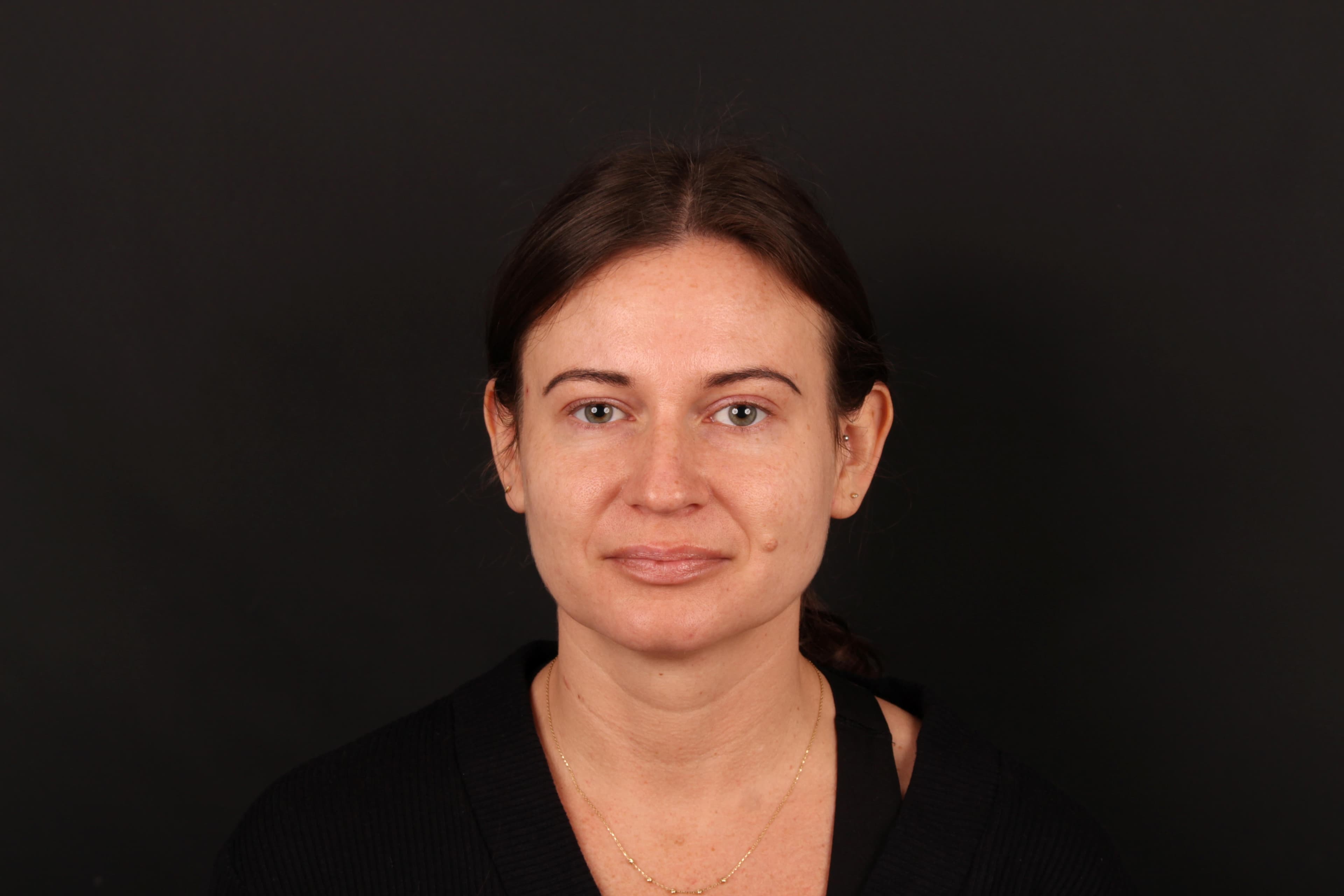
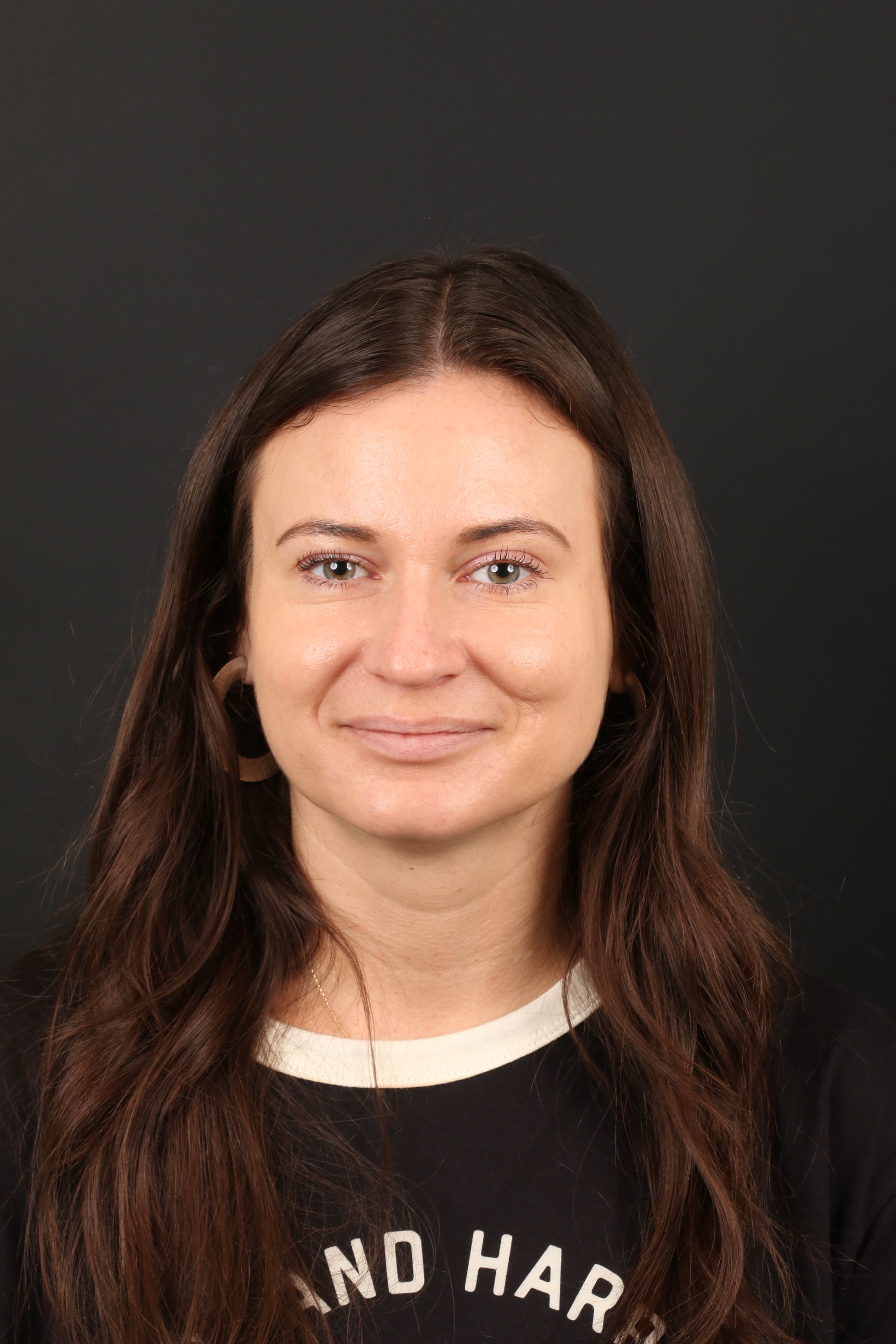
Actual patient shown before treatment and at four-month follow-up. Individual healing and results vary.
Treatment
Molectomy™
Area
Left cheek
Follow-up
4 months
01 | Case at a Glance
This case follows a left-cheek Molectomy™ from pre-operative movement assessment and scar-conscious planning through multilayer closure, pathology and four-month healing.
Primary Concern
Raised, pigmented cheek mole present since childhood and gradually increasing in size
Planning Focus
Facial movement, incision orientation, skin tension and protection of the nearby corner of the mouth
Closure
Multilayer plastic surgery closure
Pathology
Benign melanocytic nevus
Treatment
Molectomy™
Area
Left cheek
Age Range
Early 30s
Follow-up
4 months
The patient sought consultation for a mole on her left cheek that had been present since childhood and had gradually increased in size. She felt the mole distracted from her facial aesthetics and was particularly conscious of it in photographs.
Because the mole sat on a highly visible and mobile part of the face, the treatment plan needed to address more than removal alone. Dr. Ardesh also considered how the cheek moved, how the surrounding skin carried tension and how the repair could be positioned to preserve a natural facial appearance.
02 | Why Facial Movement Matters
Signature planning detail
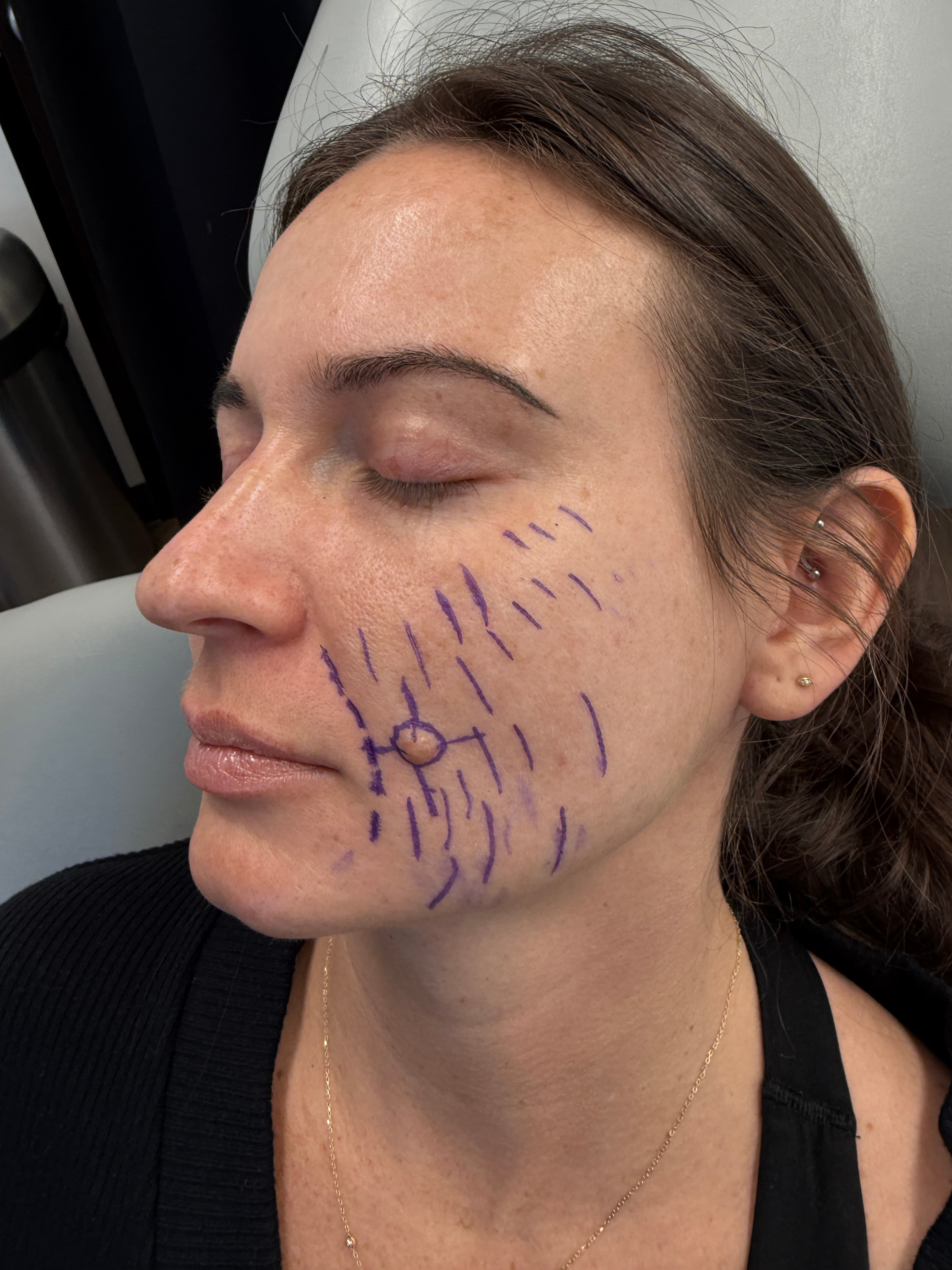
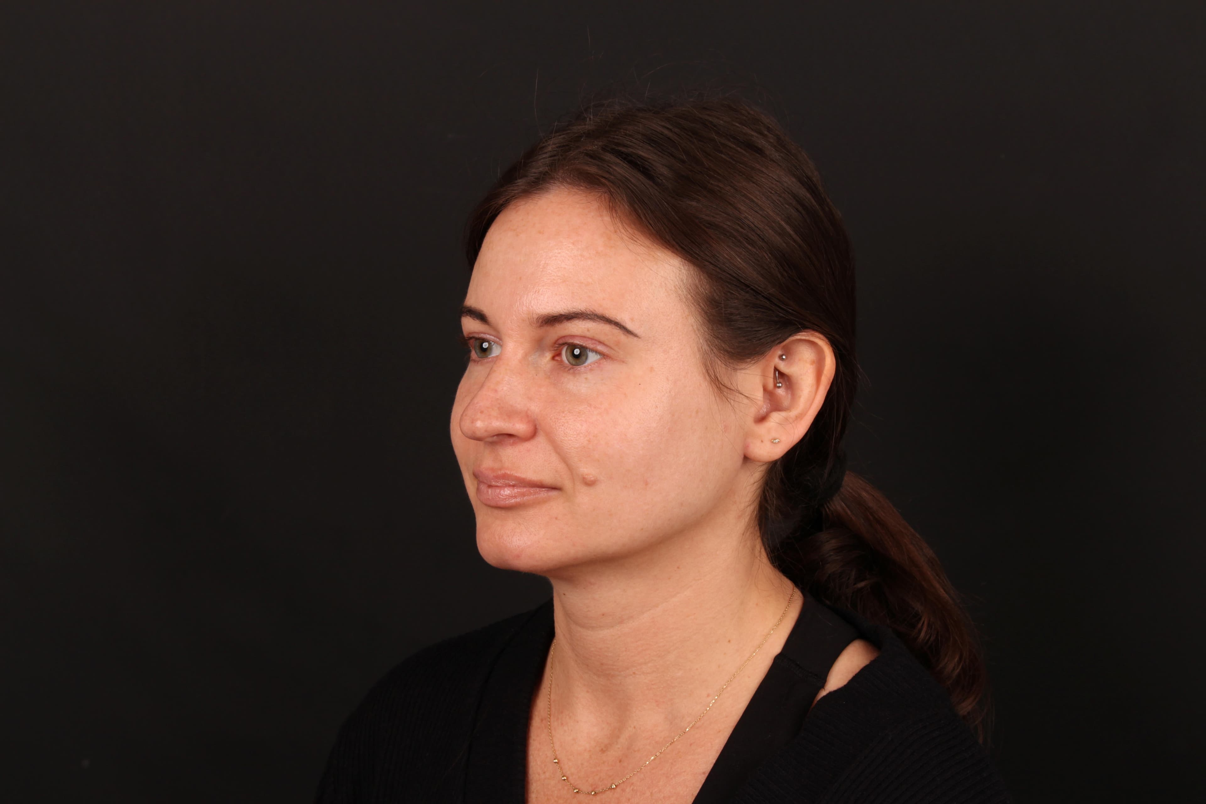
Before planning the Molectomy™, Dr. Ardesh evaluated the cheek both at rest and in motion. The patient was observed while smiling, puckering, and opening and closing her mouth.
This movement assessment helped show how the surrounding skin shifted and where tension developed. For a cheek procedure, the removal and surgical repair need to look natural when the face moves — not only when it is still.
View 01
Resting Face
View 02
Smiling
View 03
Lip Movement
View 04
Dynamic View
Why this matters
The cheek changes shape with smiling, speaking and mouth movement. Evaluating those forces before surgery helps guide incision orientation and closure planning around a mobile area of the face.
03 | Surgical Planning
Dr. Ardesh examined the mole's size, shape, projection and exact position on the cheek, together with the surrounding skin and facial contours. The planned incision was oriented to follow natural skin relaxation lines and facial contours whenever possible.
The aim was to create a repair that worked with the cheek rather than against it, while keeping the final scar as discreet and well positioned as the anatomy allowed.
Tension + mouth protection
Because the patient was relatively young with firm, tight skin, particular attention was given to tension across the repair. Excessive tension can influence how a scar heals and may contribute to widening over time.
The mole's position also required careful planning around the nearby corner of the mouth so the repair could be completed without unnecessary pulling, bunching, dog ears or visible distortion of the mouth.
04 | Molectomy™ + Multilayer Closure
Dr. Ardesh performed the cheek Molectomy™ with the goal of complete removal while preserving the natural contour of the cheek. The repair was then completed in multiple layers to support the tissues beneath the skin and reduce unnecessary surface tension.
This layered approach allowed the skin edges to be brought together carefully while keeping the cheek contour and nearby mouth anatomy in mind.
Pathology
After removal, the tissue was sent for microscopic examination by a board-certified pathologist. The pathology confirmed a benign melanocytic nevus — a noncancerous mole.
Step by step
1
Assess
Document the mole and evaluate the cheek both at rest and during facial movement.
2
Plan
Consider natural facial lines, skin tension and the nearby mouth corner before incision.
3
Remove
Perform Molectomy™ with controlled removal of the cheek mole.
4
Close
Use a multilayer plastic surgery closure to support the repair beneath the skin.
5
Review
Assess healing, facial contour and the four-month result.
05 | Four-Month Result
At the four-month follow-up, the raised, pigmented mole was no longer present. The cheek had a smooth, natural-looking contour, and the healed surgical scar blended well with the surrounding skin.
Importantly, the result maintained the natural shape of the cheek and the nearby corner of the mouth, with no noticeable distortion in the follow-up photographs.
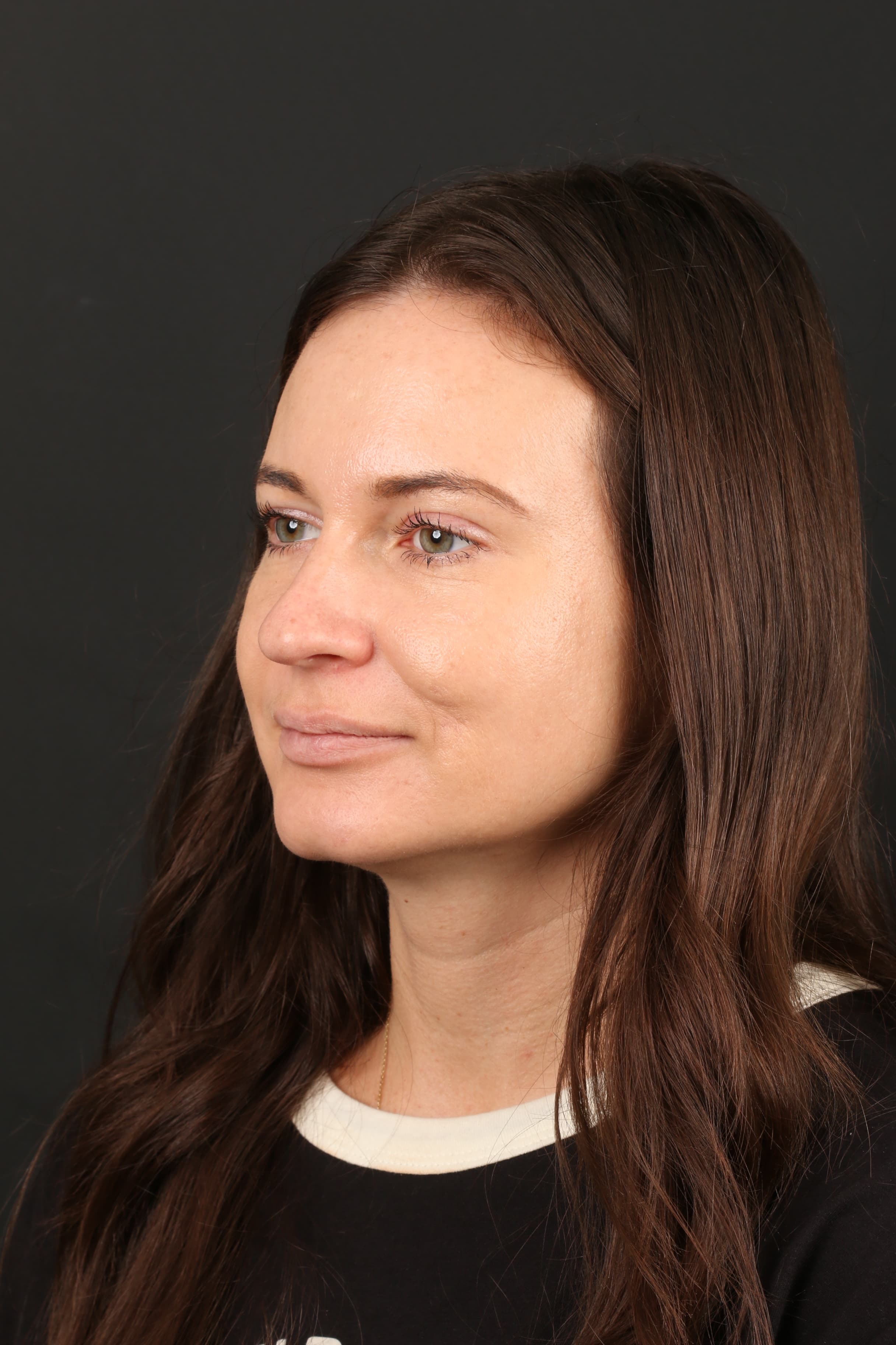
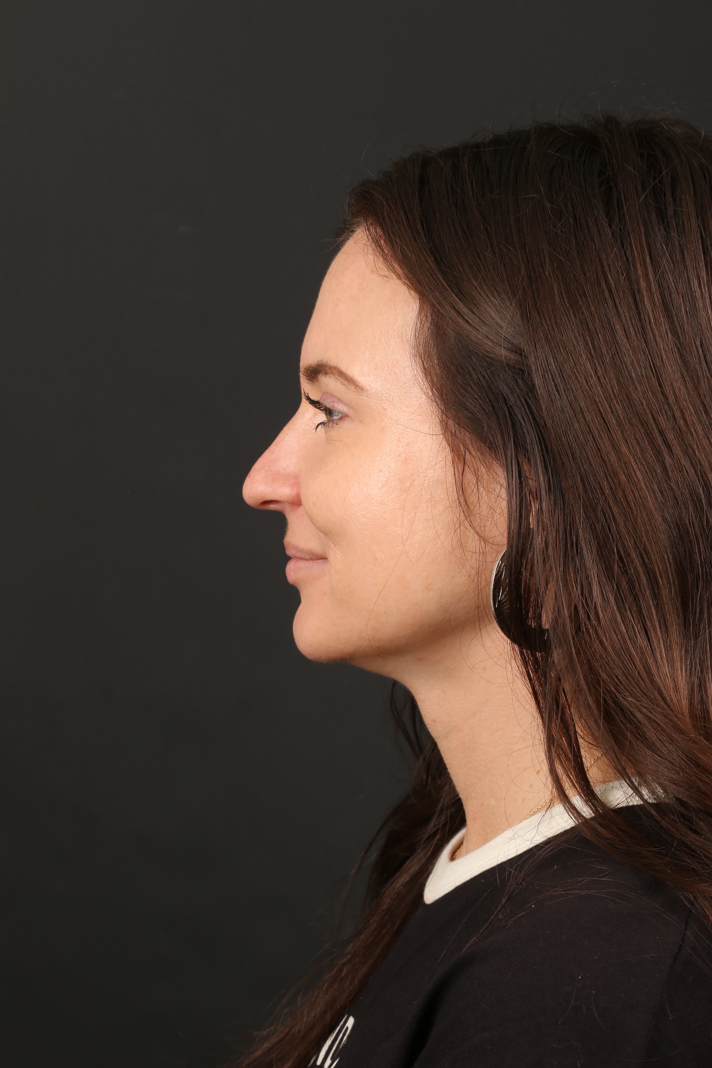
Four months after cheek Molectomy™ — healed treatment area with preserved cheek contour.
06 | Before & After
Matched or near-matched angles make it easier to compare the original cheek mole, nearby mouth contour and the healed treatment area at four months.
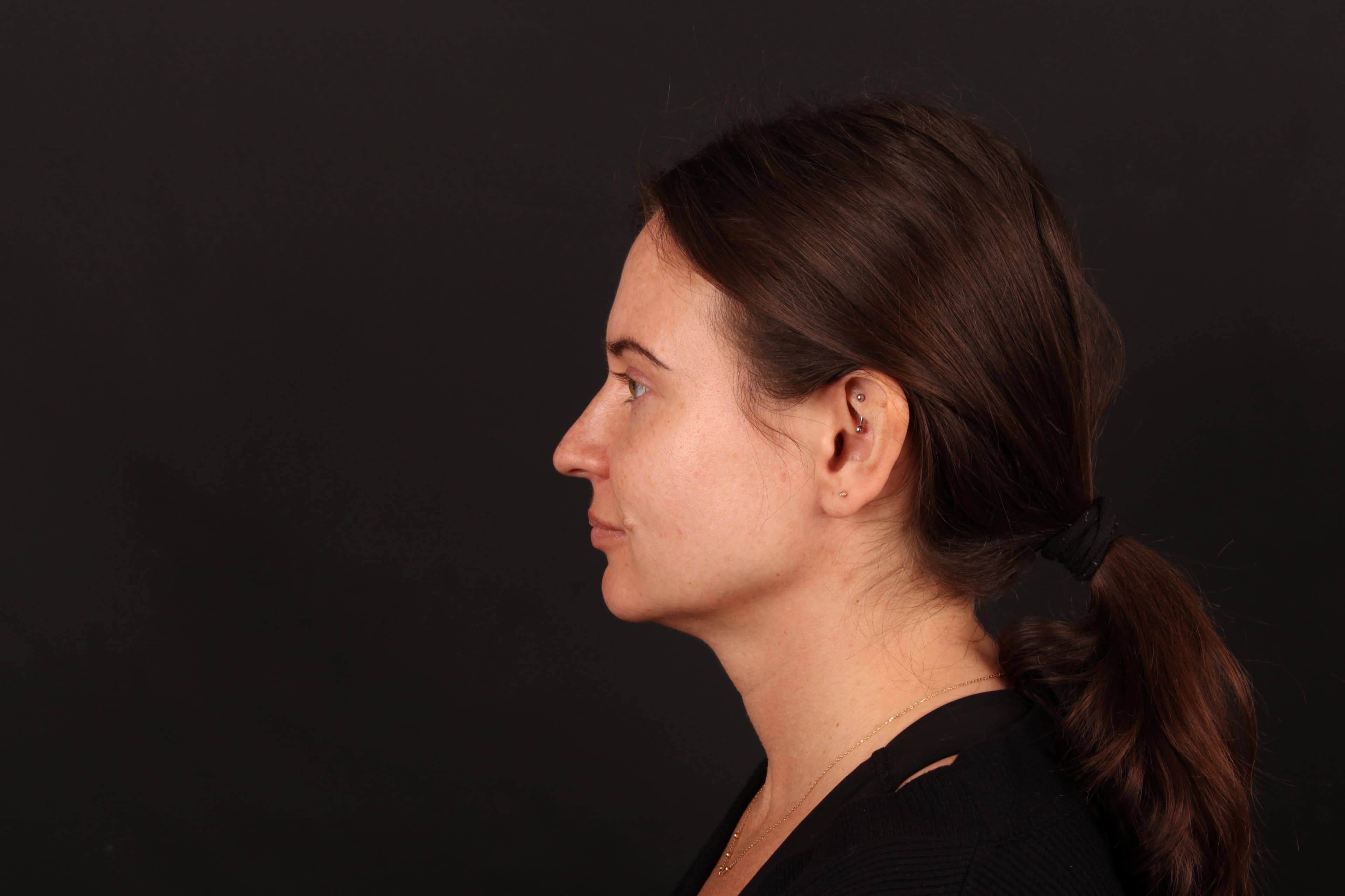
Four-Month Follow-Up
Dr. Ardesh and the patient review the four-month result, including facial movement, healing and the final cheek contour.
Additional Four-Month Follow-Up
Transformation summary
Before
Raised, pigmented cheek mole present since childhood and increasingly noticeable
Planning
Facial movement, skin tension, natural facial lines and mouth-corner position evaluated
Treatment
Molectomy™ with multilayer plastic surgery closure
Pathology
Benign melanocytic nevus
Follow-up
4 months
Outcome
Mole removed with a smooth cheek contour and a subtle healed scar blending with the surrounding skin
For a cheek mole, the surgical plan cannot be based on a still photograph alone. The cheek changes shape with smiling, speaking, puckering and mouth movement, so the incision and closure need to be planned with those forces in mind.
In this case, the position of the mole, the patient's relatively tight skin and the nearby corner of the mouth all influenced the design. The objective was complete removal with a scar-conscious repair that respected natural facial movement and contour.
Every facial mole is different. Its size, depth, location, surrounding skin, facial movement and individual healing characteristics can all influence the treatment plan. A consultation with Dr. Ardesh can help determine which mole removal approach is appropriate for your specific concern.