
1
Assess
Document the mole, its location and projection.
Case Study | Mole Removal
A 6-Month Transformation
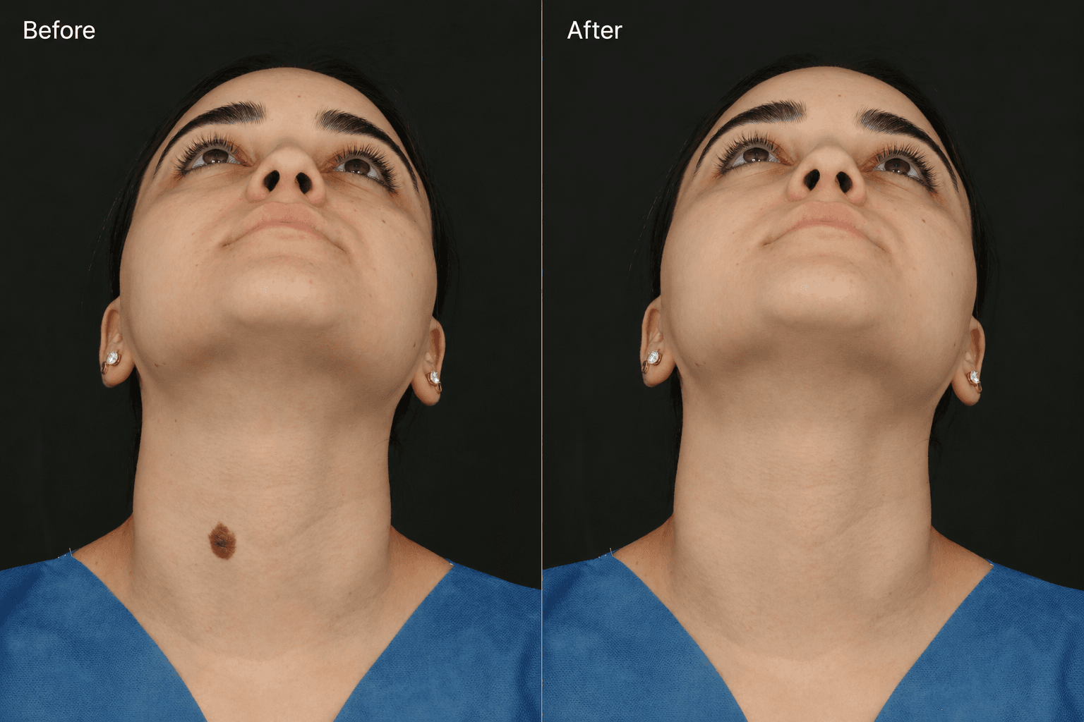
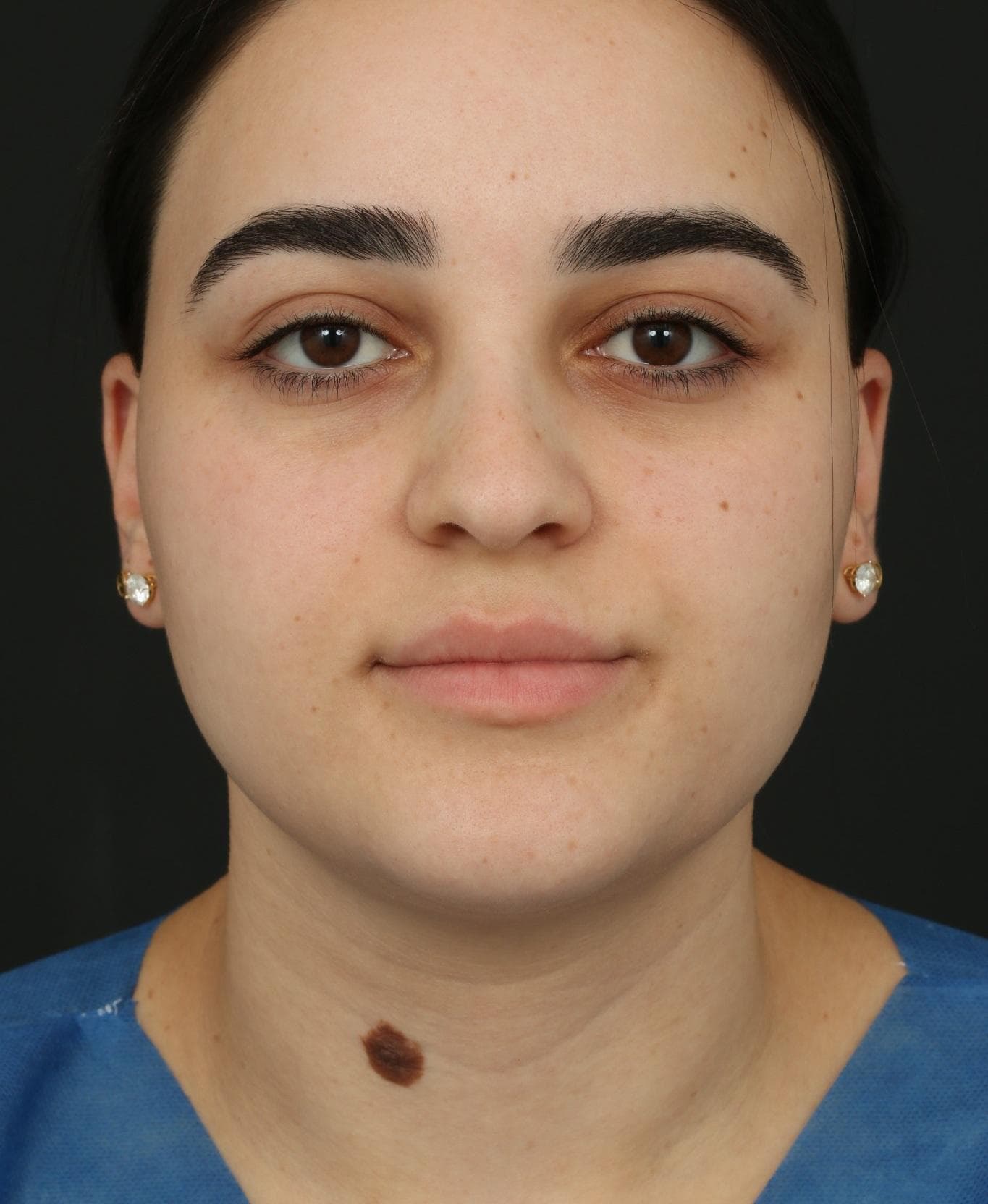
A raised, dark-brown mole on the lower neck had been present since childhood. Dr. Ardesh performed his signature Molectomy™ approach, followed by a multilayer closure with ultrafine sutures. At six months, the treatment area shows a smooth, well-healed result with a subtle scar.
Treatment
Molectomy™
Area
Lower neck
Follow-up
6 months
01 | Case at a Glance
This case documents the full treatment journey from pre-operative assessment and Molectomy™ through closure, pathology review and six-month healing.
Concern
Raised, dark-brown mole present since childhood
Treatment
Molectomy™
Closure
Multilayer closure with ultrafine sutures
Follow-up
6 months after surgery
Area
Lower neck
Removal
Underlying tissue + hair follicles
Pathology
Microscopic evaluation
Outcome
Smooth, well-healed site
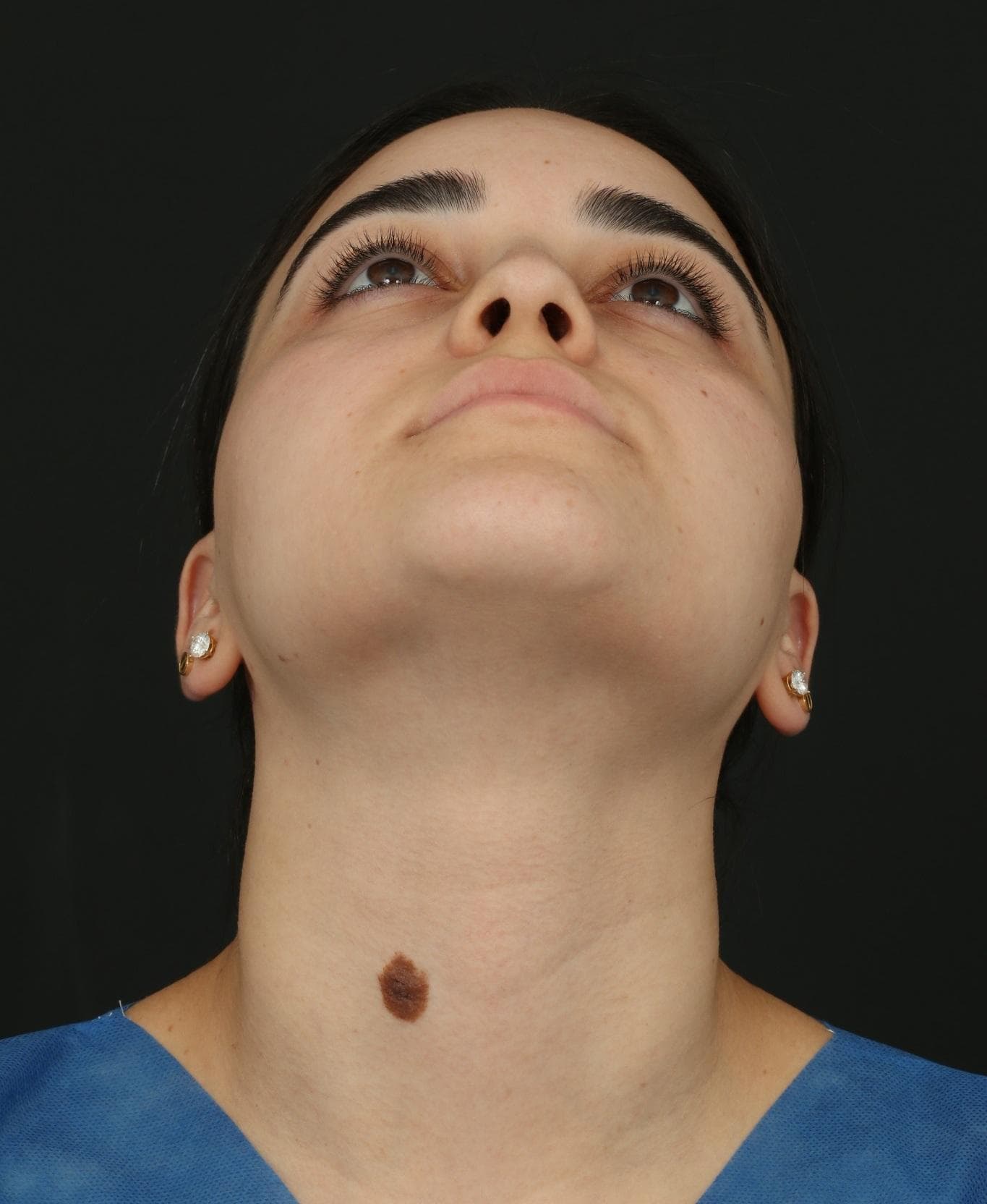
A prominent, raised and pigmented mole was visible on the lower neck. Because of its location, the treatment plan needed to consider more than removal alone. Dr. Ardesh also planned for how the area would be closed and how the resulting scar would mature over time.
02 | Assessment & Treatment Planning
Pre-treatment photography documented the mole from frontal and upward-facing views. The upward angle makes the projection, position and relationship to the surrounding neck skin especially clear, while the frontal image on the previous section provides the baseline view.
03 | The Molectomy™ Approach
Dr. Ardesh performed his signature Molectomy™ procedure to remove the visible mole together with the underlying mole tissue, including associated hair follicles. The focus was on a thorough removal while preserving a precise, controlled treatment site for closure.
Closure
Once removal was complete, Dr. Ardesh performed a multilayer closure using ultrafine sutures. This step supports accurate tissue alignment and is designed to help the incision heal as neatly as possible. For a visible area such as the neck, the quality of the closure is an important part of the overall treatment, not an afterthought.
Scar-conscious care
The case demonstrates a scar-conscious approach: careful removal, controlled tissue handling, layered closure and sufficient time for the scar to mature before judging the final appearance.
Tissue review
The removed mole was sent to a laboratory for microscopic evaluation by a board-certified pathologist. This provides a formal histologic assessment of the excised tissue after removal.
Step by step
1
Assess
Document the mole, its location and projection.
2
Remove
Perform Molectomy™ and remove the lesion and underlying tissue.
3
Close
Use multilayer closure with ultrafine sutures.
4
Review
Allow healing and assess the six-month result.
04 | Six-Month Outcome
A smooth, natural-looking result
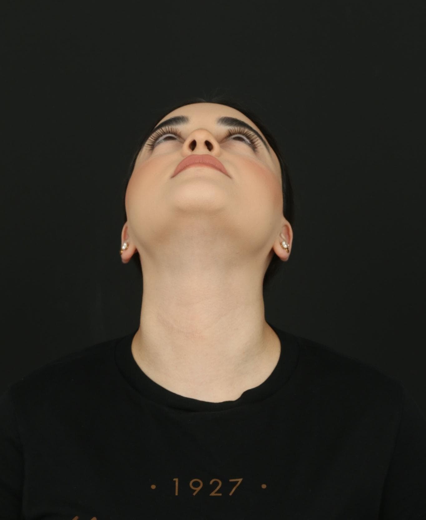
At the six-month follow-up, the mole is no longer visible and the treatment site appears flat and well healed. The scar is subtle and blends with the surrounding neck skin, demonstrating how the area settled after surgery and scar maturation.
05 | Before & After
Compare the documented pre-treatment photograph with the six-month follow-up result.
Actual treatment images shown before surgery and at six-month follow-up. Individual healing and results vary.
This case illustrates that mole removal is not simply about taking away what is visible on the skin. Assessment, depth of removal, tissue handling, closure technique, pathology review and follow-up all contribute to the quality of the final result.
Every mole and every treatment area is different. A consultation allows Dr. Ardesh to assess the lesion, discuss the most appropriate removal approach and explain what to expect from healing and scar maturation.