
1
Assess
Evaluate the number, location, projection and spacing of the cheek moles.
Mole Removal • Transformation Story
An early follow-up transformation focused on complete removal, facial-movement planning, refined closure and natural-looking healing.
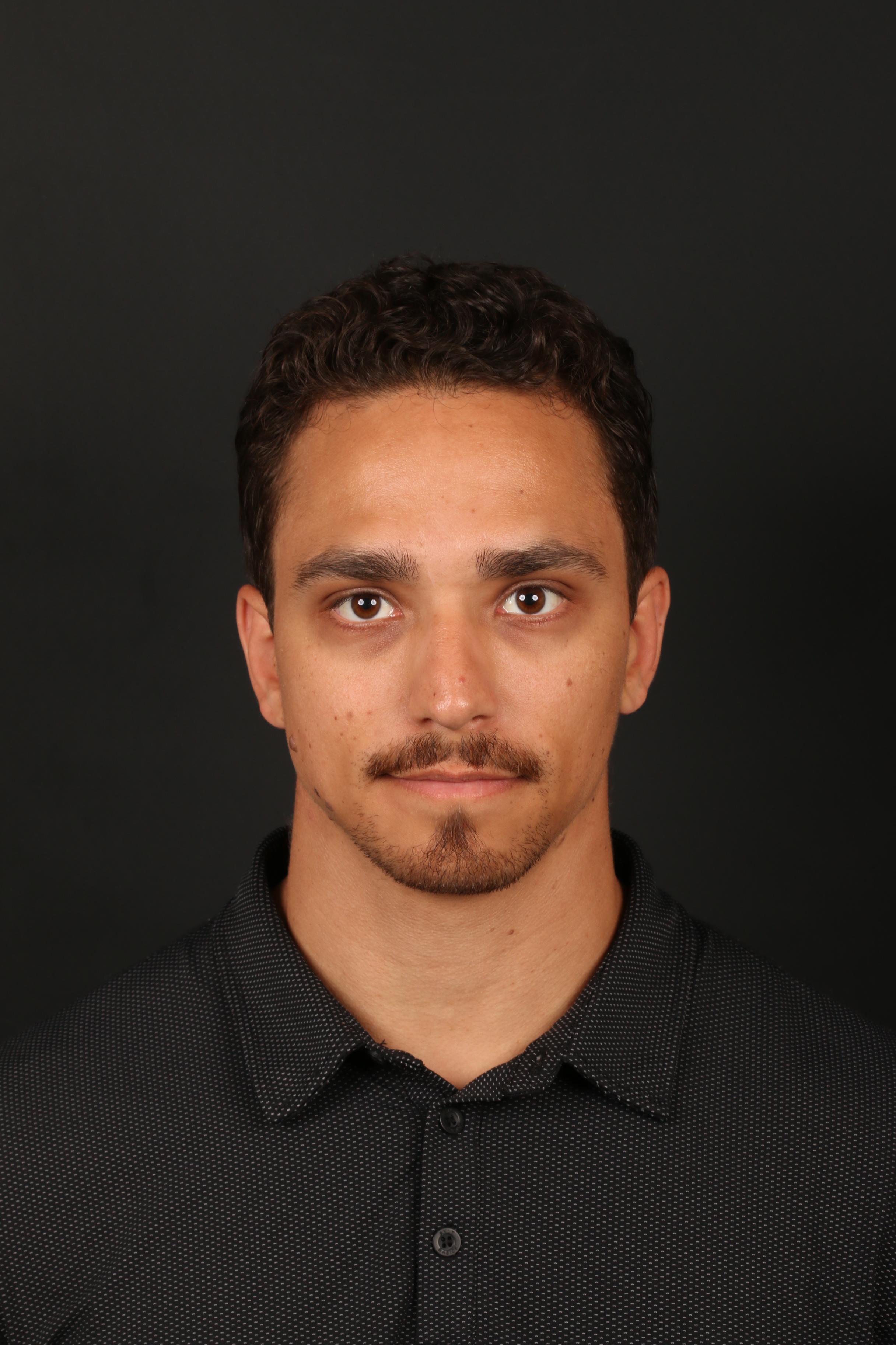
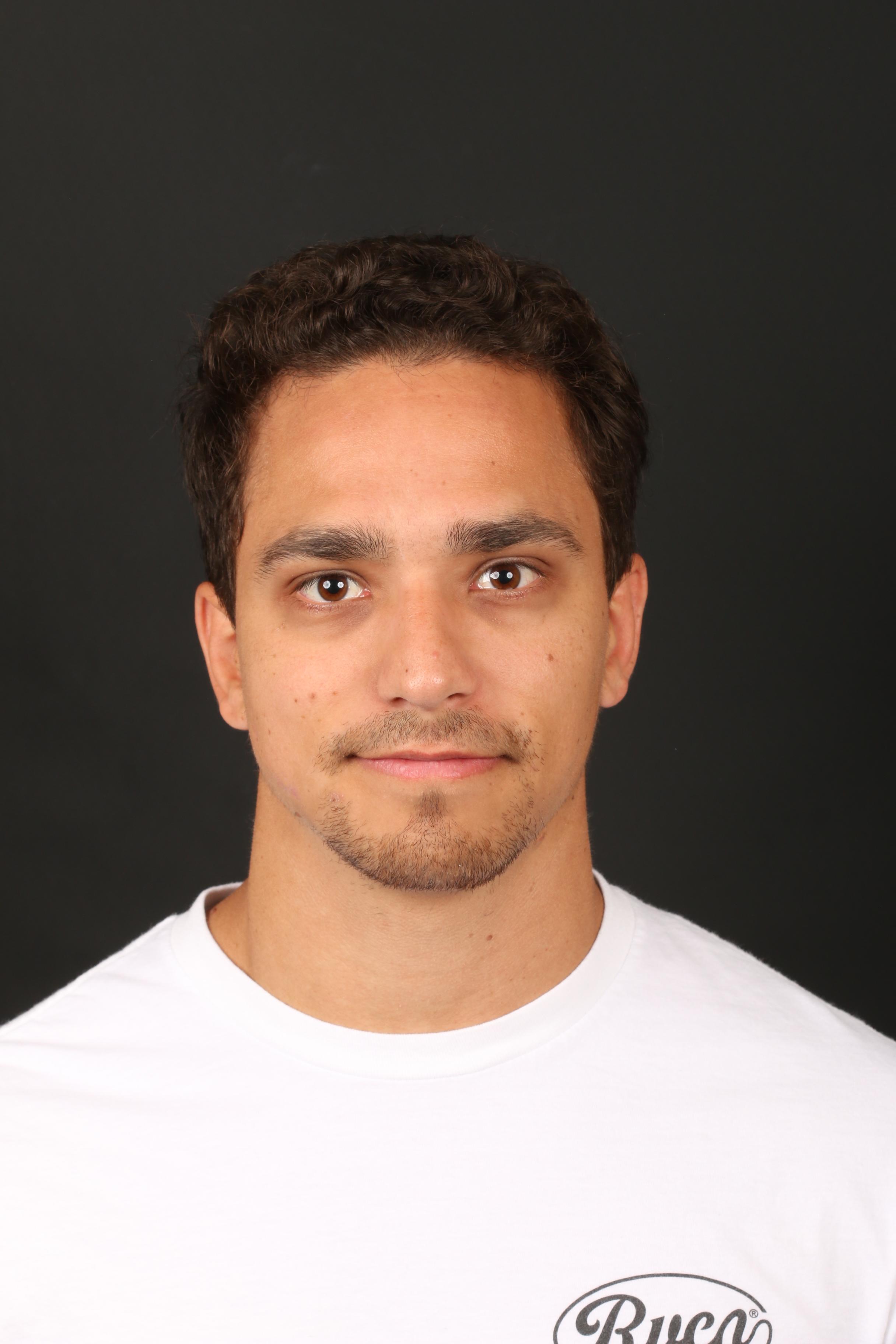
Actual patient. Individual healing and results vary.
Treatment
Molectomy™
Area
Cheek
Follow-up
Early follow-up
01 | Case at a Glance
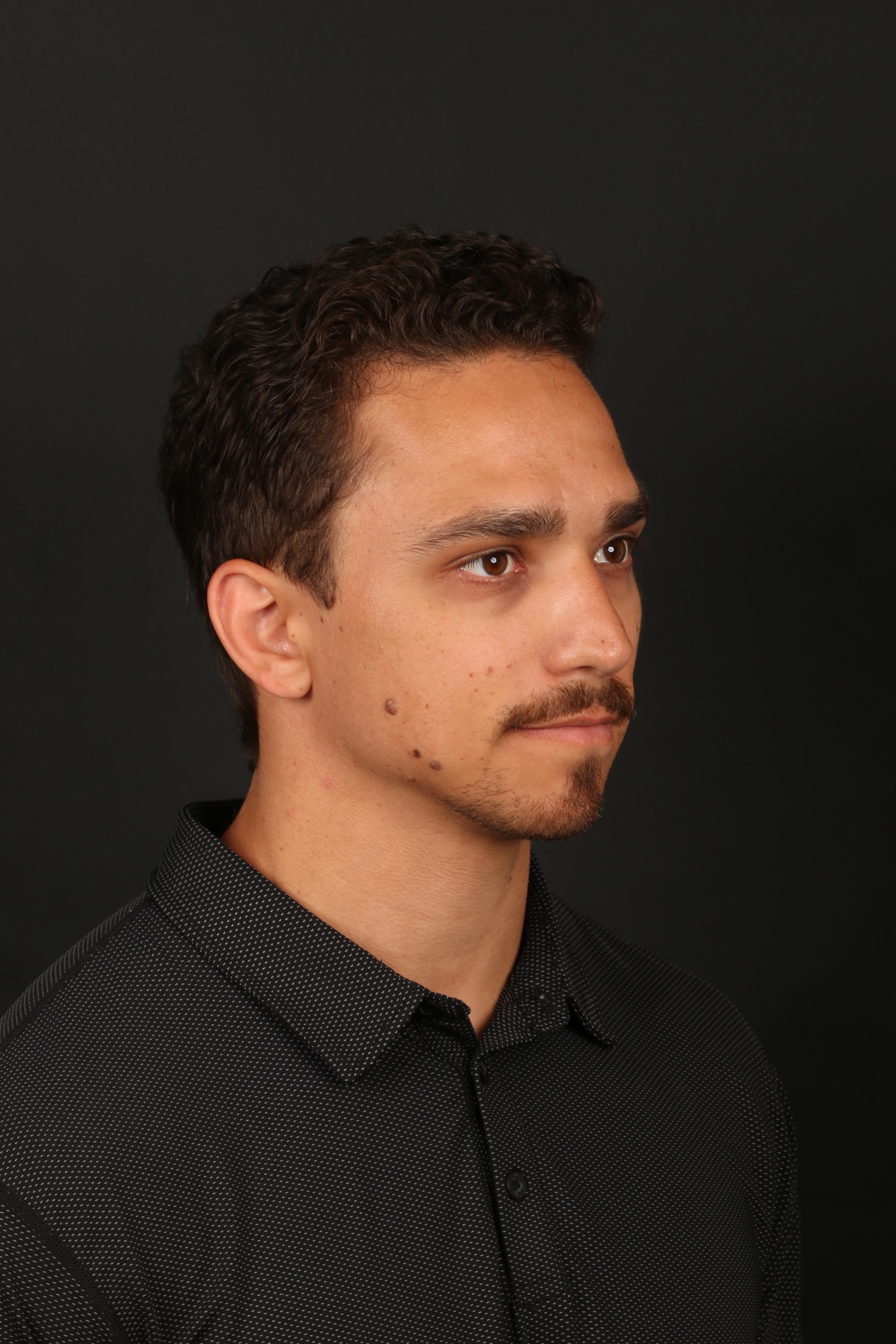
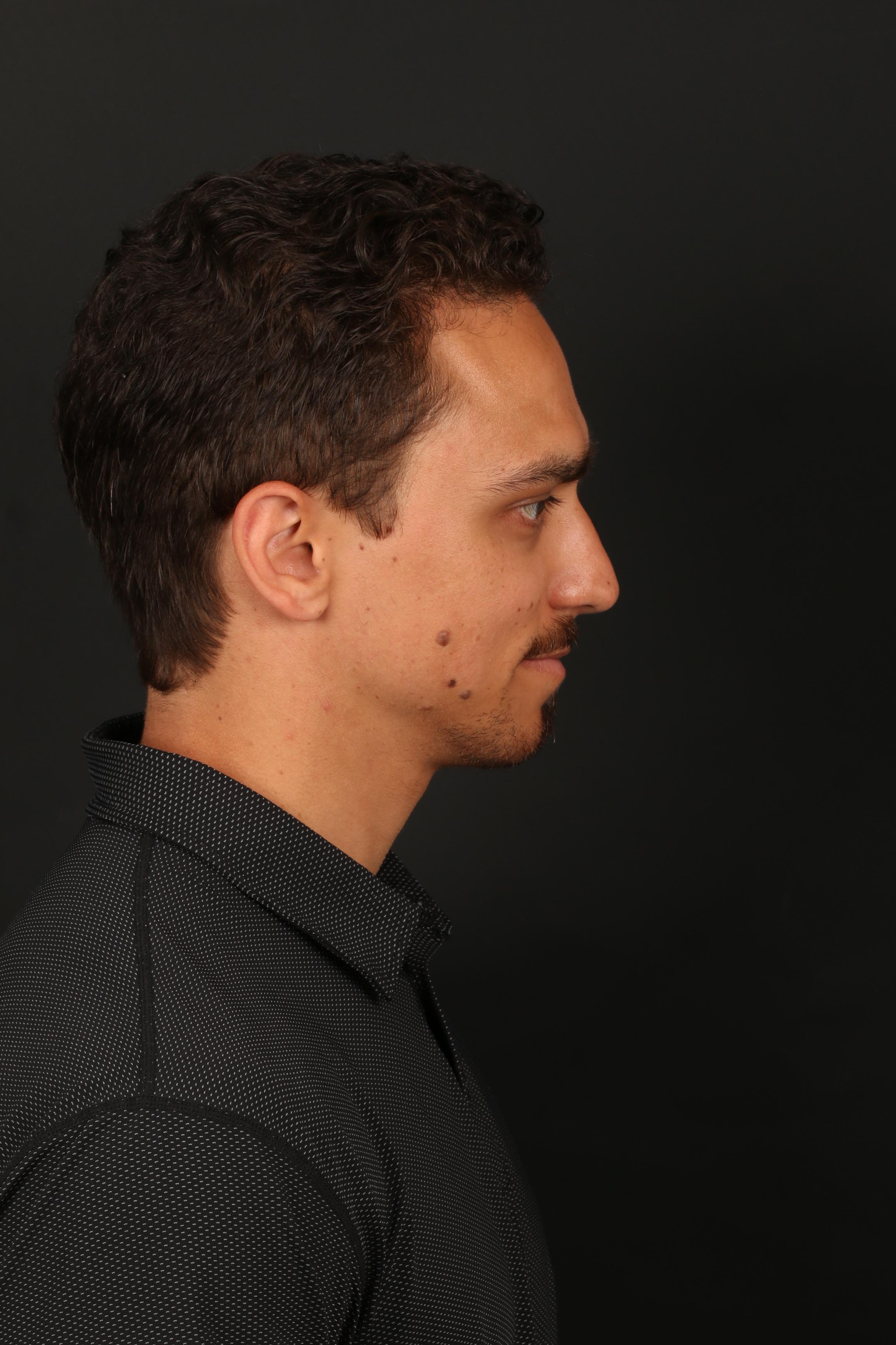
This 31-year-old man wanted removal of several raised, dark-brown moles on his cheek. Because multiple lesions were present across the same facial area, the treatment plan needed to consider not only each mole individually, but also how the separate surgical sites would heal together within the natural contour of the cheek.
The objective was complete removal of each mole while keeping the final scars as refined and unobtrusive as possible.
Primary Concern
Several raised, dark-brown cheek moles
Planning Focus
Facial movement, scar placement and a coordinated closure strategy
Closure
Multilayer closure with ultrafine sutures
Pathology
Benign nevi
Treatment
Molectomy™
Area Treated
Cheek
Patient
31-year-old man
Result
Natural-looking cheek contour
Several individual treatment sites meant the plan had to account for scar direction, spacing and how the cheek would move as a whole after repair.
02 | Assessment & Facial Movement
Movement-based planning
Dr. Ardesh evaluated the location, projection and spacing of the cheek moles and also considered normal facial movement, including smiling. The cheek changes shape and tension with expression, so the direction and strength of each closure need to work with that movement rather than against it.
Why this matters
With several moles in the same region, planning is not just about individual excisions. The relationship between the separate closures, facial expression and overall cheek contour matters to the final result.
03 | The Molectomy™ Approach
Dr. Ardesh performed his signature Molectomy™ to completely remove each cheek mole, including the deeper mole components and associated hair follicles. With several treatment sites in one area, each excision and repair had to be planned carefully so the cheek maintained a natural overall appearance.
Why multiple sites require coordination
The goal is not simply to remove several separate lesions. The final result depends on how the individual closures relate to the cheek as a whole, including skin tension, expression and scar direction.
1
Assess
Evaluate the number, location, projection and spacing of the cheek moles.
2
Plan
Account for resting contour, smiling movement, scar direction and closure relationships.
3
Remove
Perform Molectomy™ for complete removal of each mole, including deeper components and associated hair follicles.
4
Repair
Close each site with a multilayer repair using ultrafine sutures and deeper support.
5
Review
Assess healing, contour and scar maturation across all treated sites at follow-up.
04 | Multilayer Closure
After removal, each site was repaired with a multilayer closure using ultrafine sutures. A layered repair provides support beneath the skin while allowing the surface edges to come together precisely.
This approach is particularly important on the cheek, where the skin moves repeatedly with expression and where a well-supported closure can help reduce unnecessary surface tension during healing.
01
Multilayer repair supports the closure beneath the skin rather than relying on the surface alone.
02
Ultrafine sutures allow the surface edges to come together with refined alignment.
03
The repair is planned for a cheek that repeatedly changes tension with facial expression.
05 | Pathology
The removed moles were sent to a laboratory for microscopic evaluation by a board-certified pathologist. The specimens were confirmed to be benign nevi.
Pathology result
Benign nevi
Confirmed on microscopic examination.
06 | Follow-Up Result
At the documented early follow-up, the treated cheek showed excellent healing. The raised moles were no longer present, and the cheek maintained a smooth, natural-looking contour across the treated areas.
The multiple small scars were continuing to mature, demonstrating the value of precise removal, thoughtful scar placement and carefully supported closure.
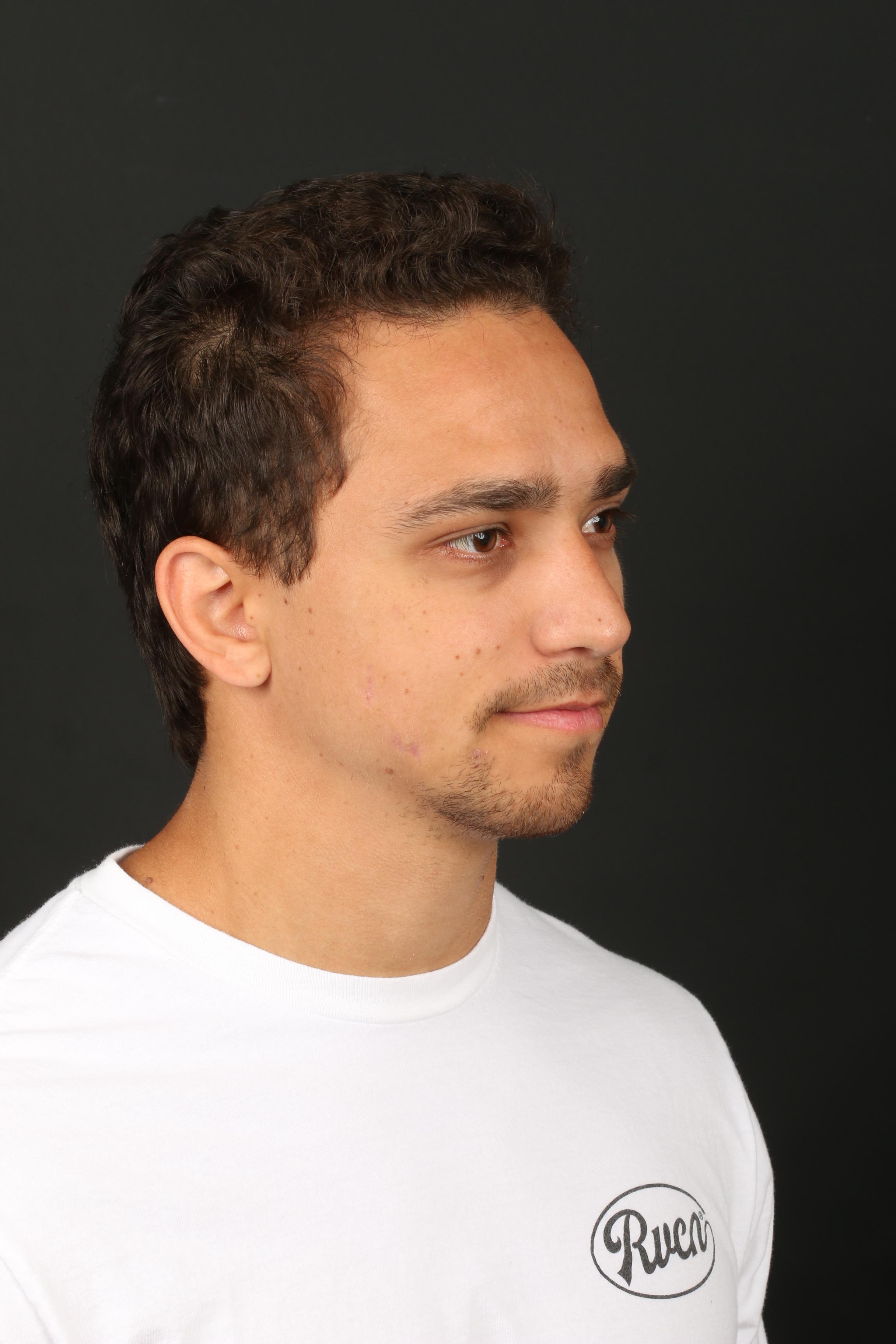
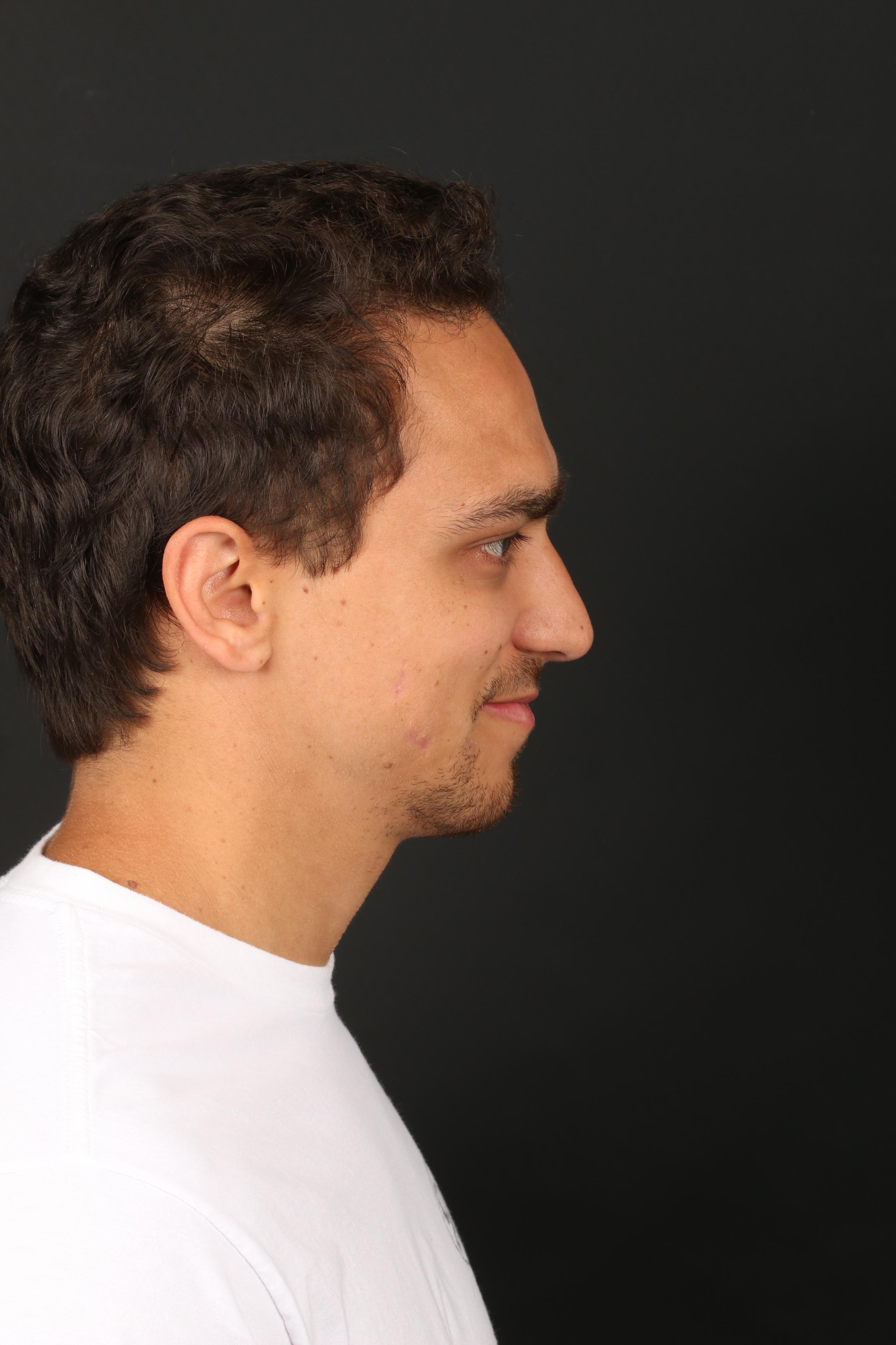
Follow-up timing should be confirmed before publishing. Individual healing and results vary.
07 | Before & After
Matched front, three-quarter and profile views make it easier to compare the treated cheek without relying on different camera angles.
Transformation summary
Before
Several raised, dark-brown cheek moles
Planning
Resting and smiling facial movement considered
Procedure
Molectomy™ for complete removal of each mole
Closure
Multilayer repair with ultrafine sutures
Pathology
Benign nevi
Result
Excellent early healing with a natural-looking cheek contour
In this case, complete removal was only one part of the treatment plan. Several individual surgical sites had to be considered together so that scar direction, skin tension, facial expression and cheek contour remained balanced across the treated area.
The objective was precise removal of each mole with supported closures that could heal naturally within a highly visible and mobile facial region.
Every mole and every treatment area is different. A consultation with Dr. Ardesh can help determine the most appropriate treatment approach based on the characteristics of the moles, their location, surrounding facial movement and individual treatment goals.