
1
Assess
Document the mole and its relationship to the nasal contours.
Case Study | Mole Removal
A 6-Month Transformation
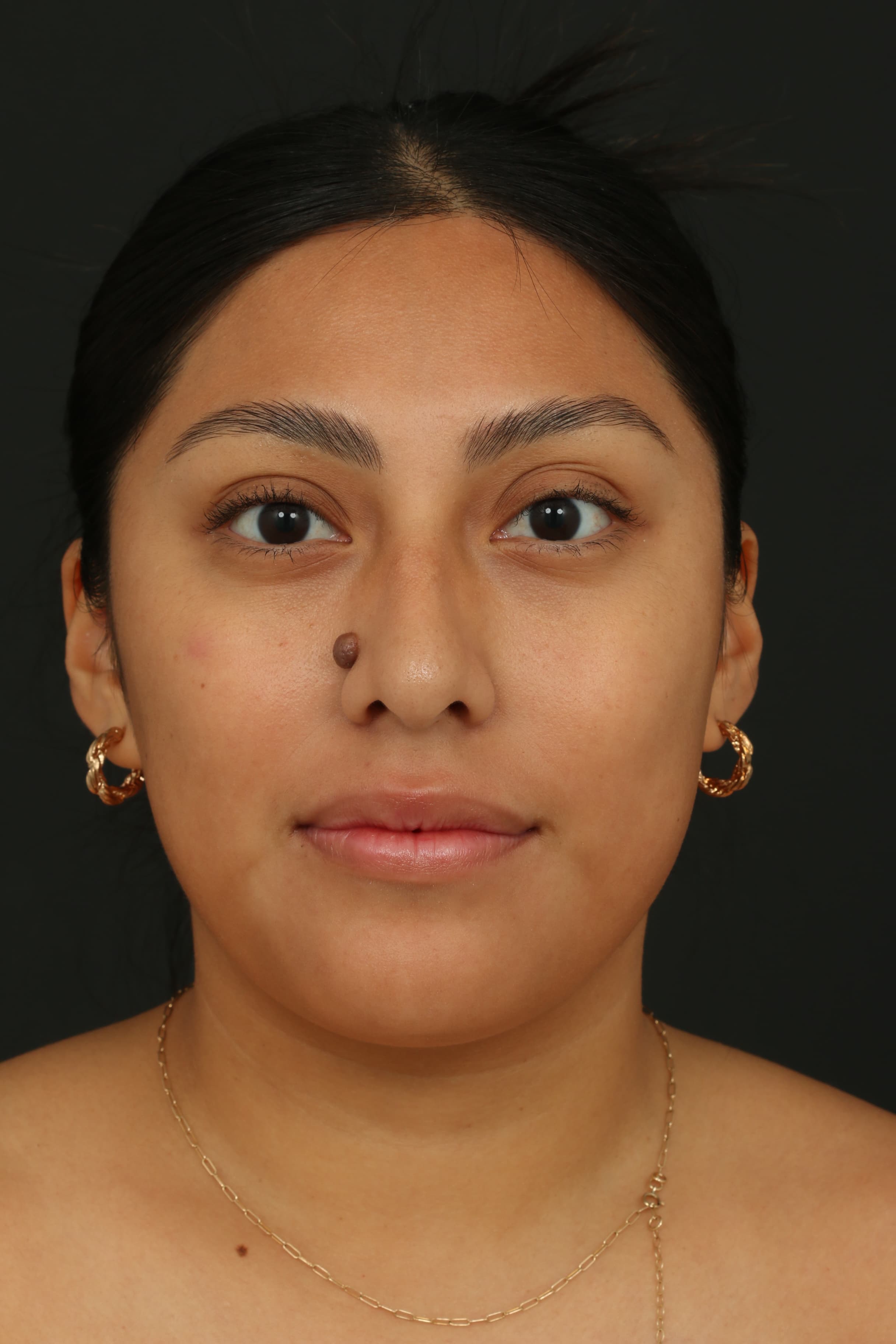
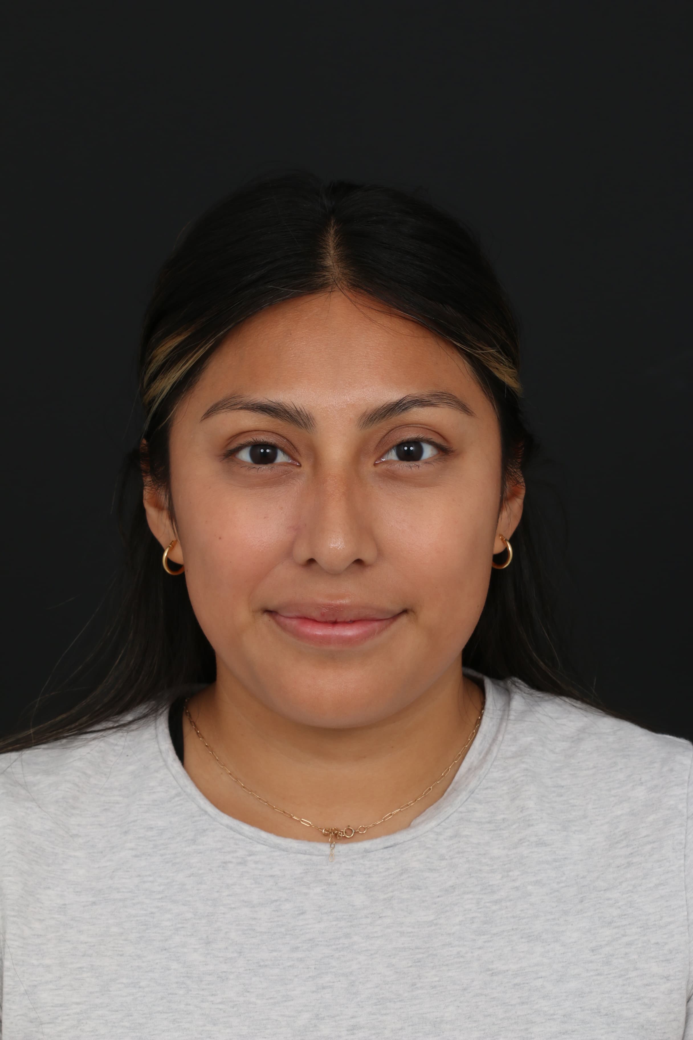
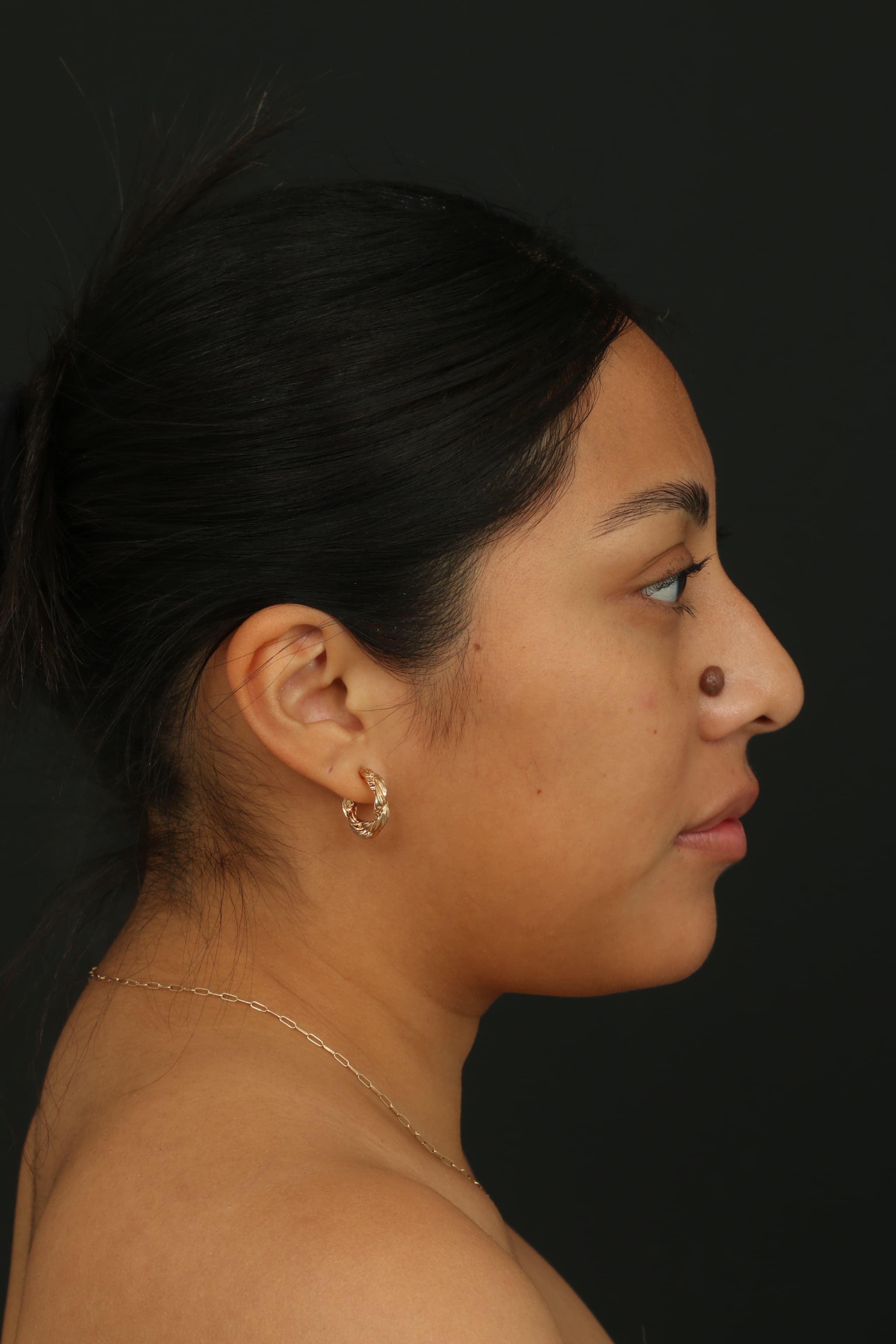
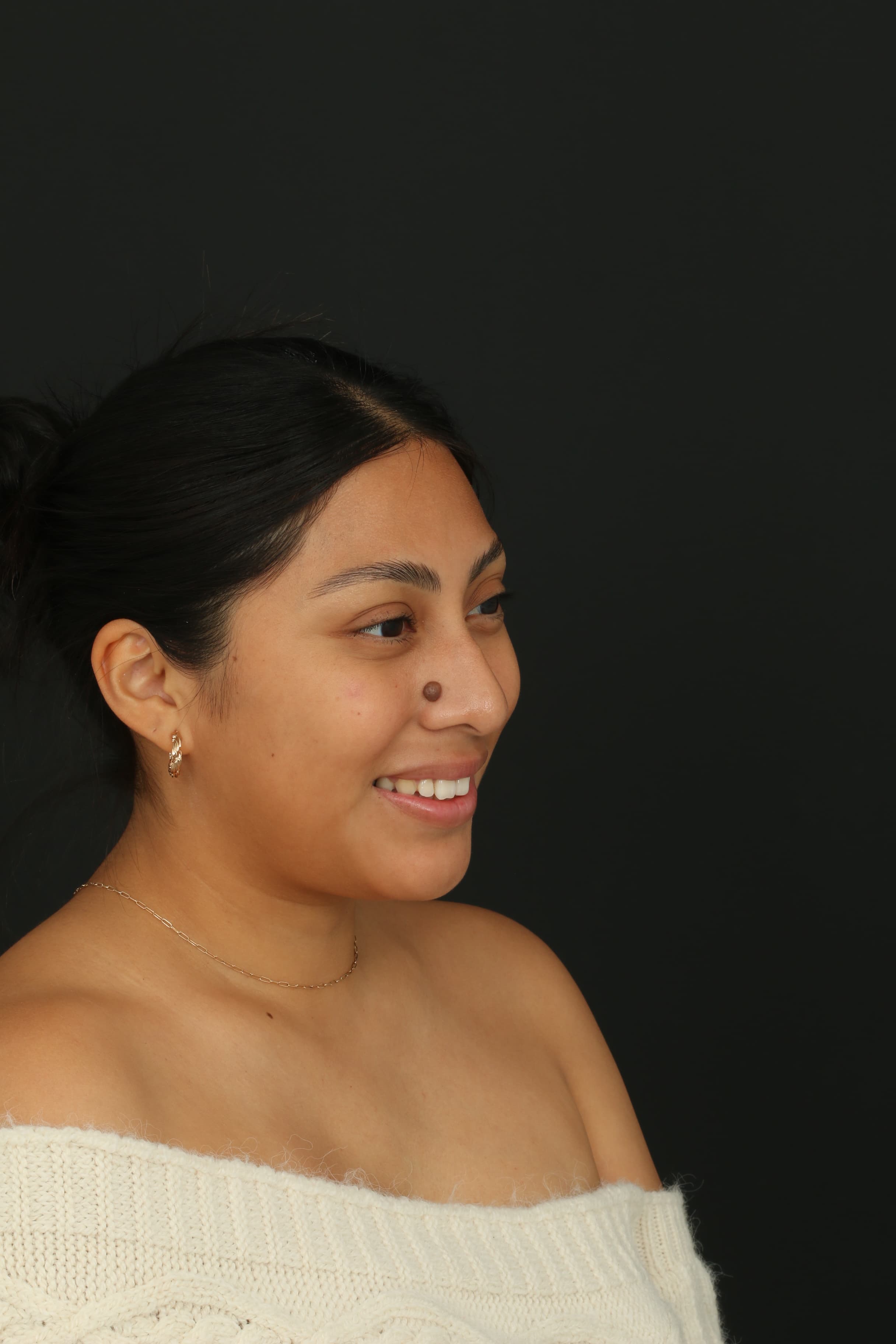
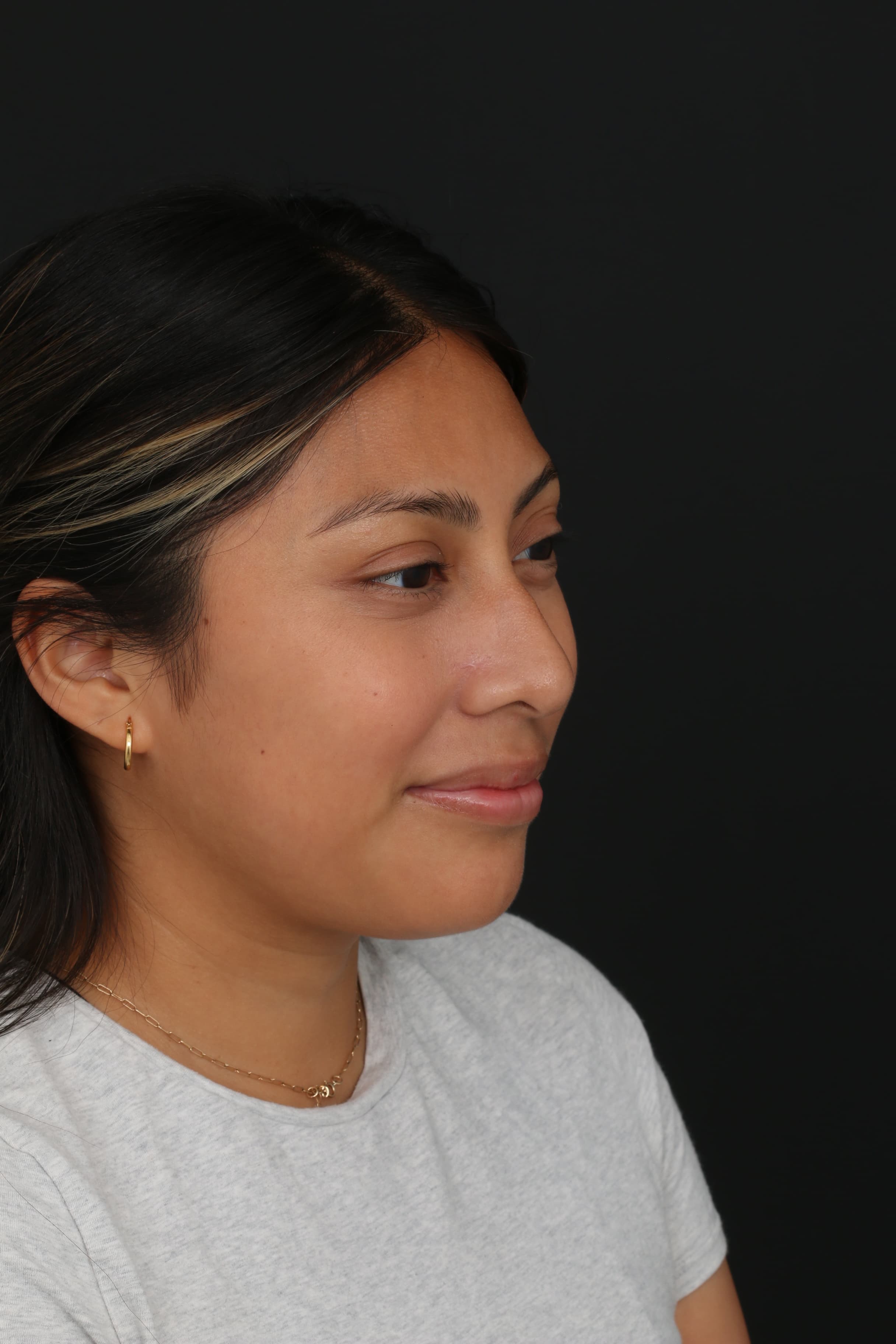
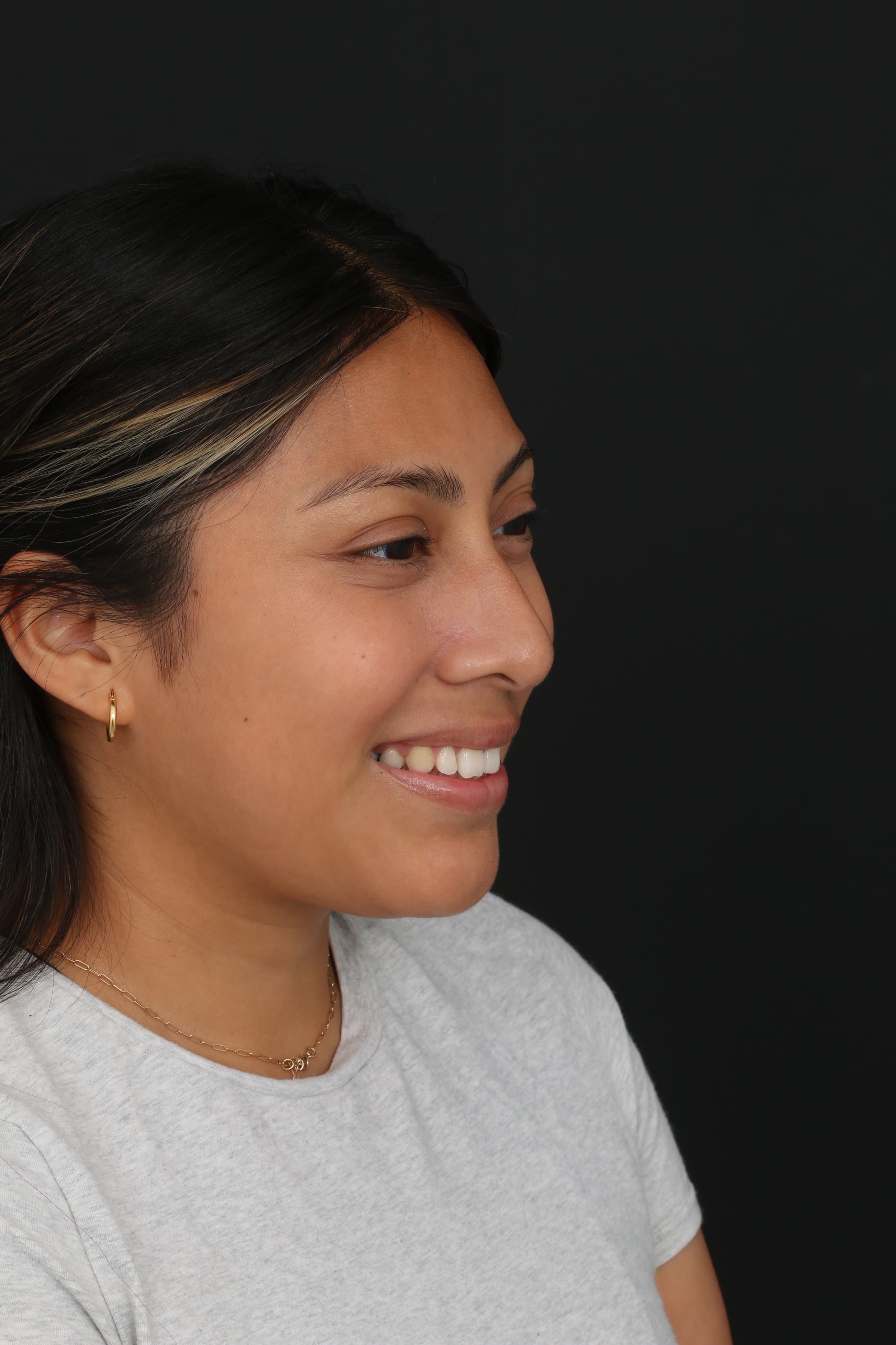
A large, raised, dark-brown mole on the nose had been present since childhood. Dr. Ardesh performed his signature Molectomy™, using careful nasal subunit planning and a multilayer closure with ultrafine sutures. At six months, the mole is no longer present and the treatment area shows a natural-looking healed result.
Treatment
Molectomy™
Area
Nose
Follow-up
6 months
01 | Case at a Glance
This case documents the treatment journey from pre-operative assessment and nasal planning through Molectomy™, multilayer closure, pathology submission and six-month healing.
Concern
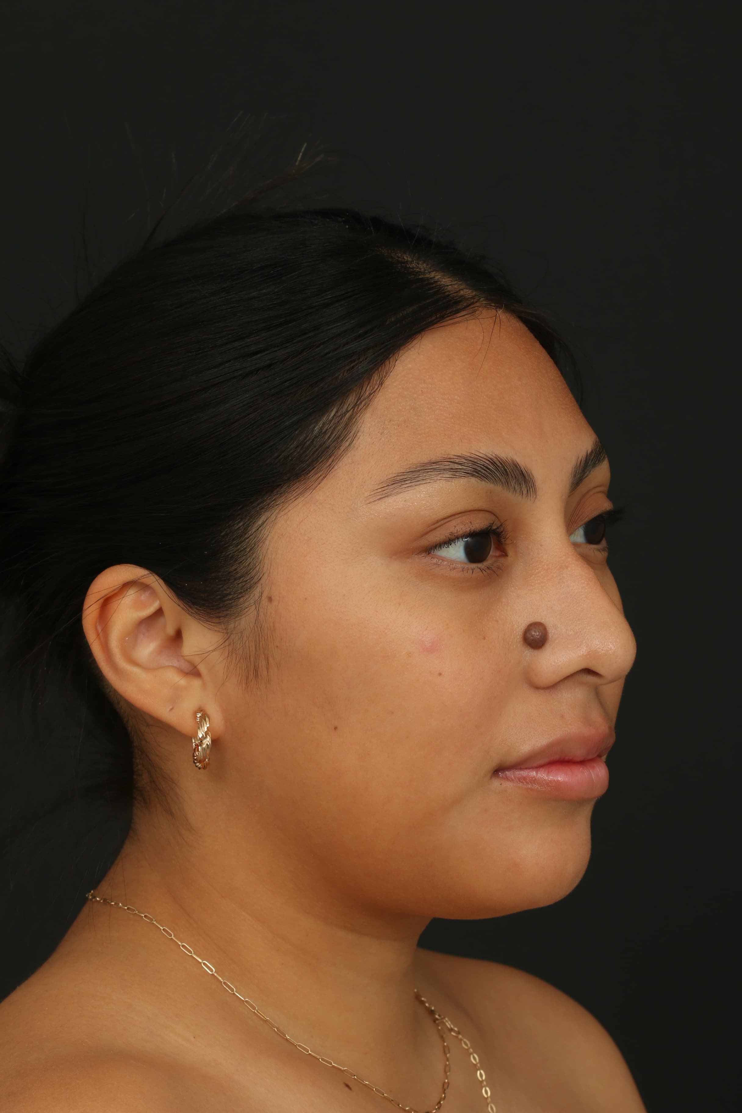
Large, raised, dark-brown mole present since childhood
Treatment
Molectomy™
Planning
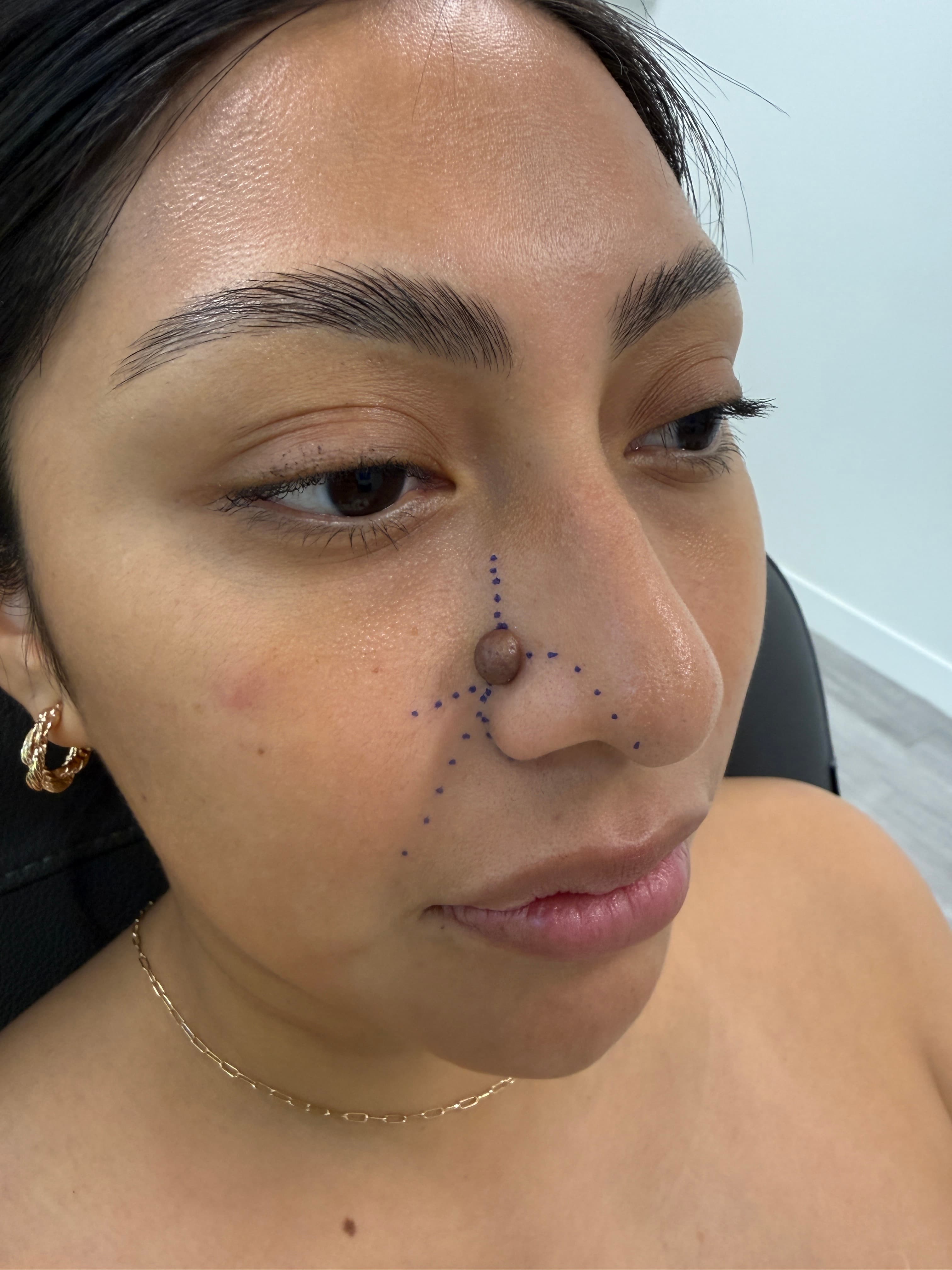
Nasal subunit markings and contour-aware incision planning
Closure
Multilayer closure with ultrafine sutures
Area
Nose
Follow-up
6 months after surgery
Pathology
Microscopic examination
Goal
Natural-looking healed result
The patient wanted removal of a prominent mole on the nose that had been present since childhood. Its size, projection and dark pigmentation made it especially noticeable in photographs. Because the nose is a central facial feature with defined contours and relatively limited skin mobility, treatment planning needed to consider both complete removal and how the incision would heal within the surrounding nasal anatomy.
02 | Assessment & Treatment Planning
Before surgery, Dr. Ardesh evaluated the mole in relation to the surrounding nasal contours and marked the planned treatment area. Nasal subunit planning helps organize the incision around the natural architecture of the nose rather than treating the mole as an isolated surface spot.
On the nose, even a small change in scar position or tissue tension can be visually noticeable. The treatment plan therefore considered the mole's location, the direction of the planned incision, the available surrounding skin and the need to avoid unnecessary distortion of the nasal contour.
03 | The Molectomy™ Approach
Dr. Ardesh performed Molectomy™ to remove the visible mole and the underlying mole tissue in a controlled surgical excision. The procedure was planned with the contours of the nose in mind so the treatment site could be repaired precisely after removal.
Closure
After the mole was removed, Dr. Ardesh performed a multilayer closure using ultrafine sutures. This approach supports accurate tissue alignment and distributes tension through deeper layers rather than relying only on the skin surface.
Tissue review
The removed tissue was sent to a laboratory for microscopic examination by a board-certified pathologist. The case summary confirms submission for pathology review; the final pathology diagnosis should only be added if the approved report is available.
Step by step
1
Assess
Document the mole and its relationship to the nasal contours.
2
Plan
Mark the nasal subunits and determine the direction of excision and closure.
3
Remove
Perform Molectomy™ and remove the lesion in a controlled fashion.
4
Close
Use a multilayer repair with ultrafine sutures.
5
Review
Allow healing and assess the six-month result.
04 | Six-Month Outcome
A natural-looking healed result
At the six-month follow-up, the raised pigmented mole is no longer present. The nasal contour remains natural, and the healed treatment site is subtle in standard frontal and three-quarter views. The result demonstrates how the treatment area settled after surgery and scar maturation.
05 | Before & After
Matching or near-matching angles help compare the mole, nasal contour and healed treatment site without distraction from different camera positions.
Six-month clinical commentary covering nasal subunit planning, scar placement, closure technique and final healing.
Patient experience and six-month result discussion.
This case illustrates why nose mole removal involves more than removing what is visible on the surface. The location of the mole, nasal subunit anatomy, direction of closure, tissue tension, fine suture technique, pathology review and adequate healing time all contribute to the final result.
Every mole and every treatment area is different. A consultation allows Dr. Ardesh to assess the lesion, discuss the most appropriate removal approach and explain what to expect from healing and scar maturation.