
01
Natural Crease Placement
Plan the repair in relation to eyelid folds so the scar can settle more discreetly.
Mole Removal • Transformation Story
A four-month transformation focused on natural crease placement, refined closure and smooth healing near the medial canthus.


Actual patient shown before treatment and at four-month follow-up. Individual healing and results vary.
Treatment
Molectomy™
Area
Upper eyelid / medial canthus
Follow-up
4 months
01 | Case at a Glance
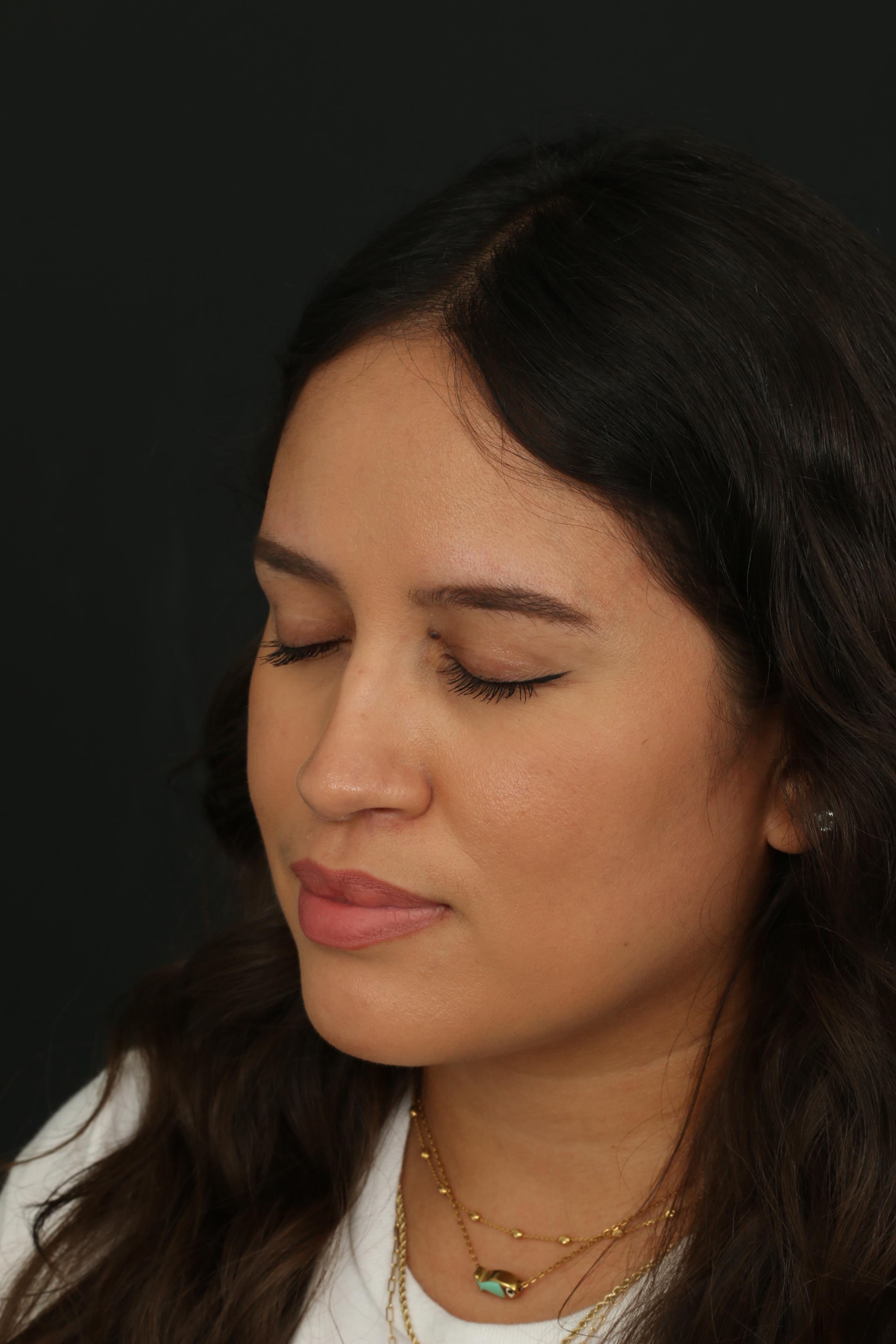
A raised, pigmented mole near the upper eyelid and medial canthus had been present since childhood and had gradually increased in size. It had become especially noticeable in photographs, prompting the patient to seek removal.
Because this area sits beside the inner corner of the eye, treatment required careful planning around eyelid movement, natural skin folds, scar direction and the risk of webbing or bunching.
Primary Concern
Raised pigmented mole present since childhood
Planning Focus
Natural crease placement, smooth eyelid movement and webbing prevention
Pathology
Benign melanocytic nevus
Follow-up
4 months
Treatment
Molectomy™
Area Treated
Upper eyelid / medial canthus
Repair Focus
Refined eyelid-specific closure
Result
Smooth, natural-looking contour
The patient felt the mole distracted from her facial aesthetics, particularly in photographs. Its position near the medial canthus made the planned repair just as important as the removal itself.
02 | Assessment & Planning
Movement-based assessment
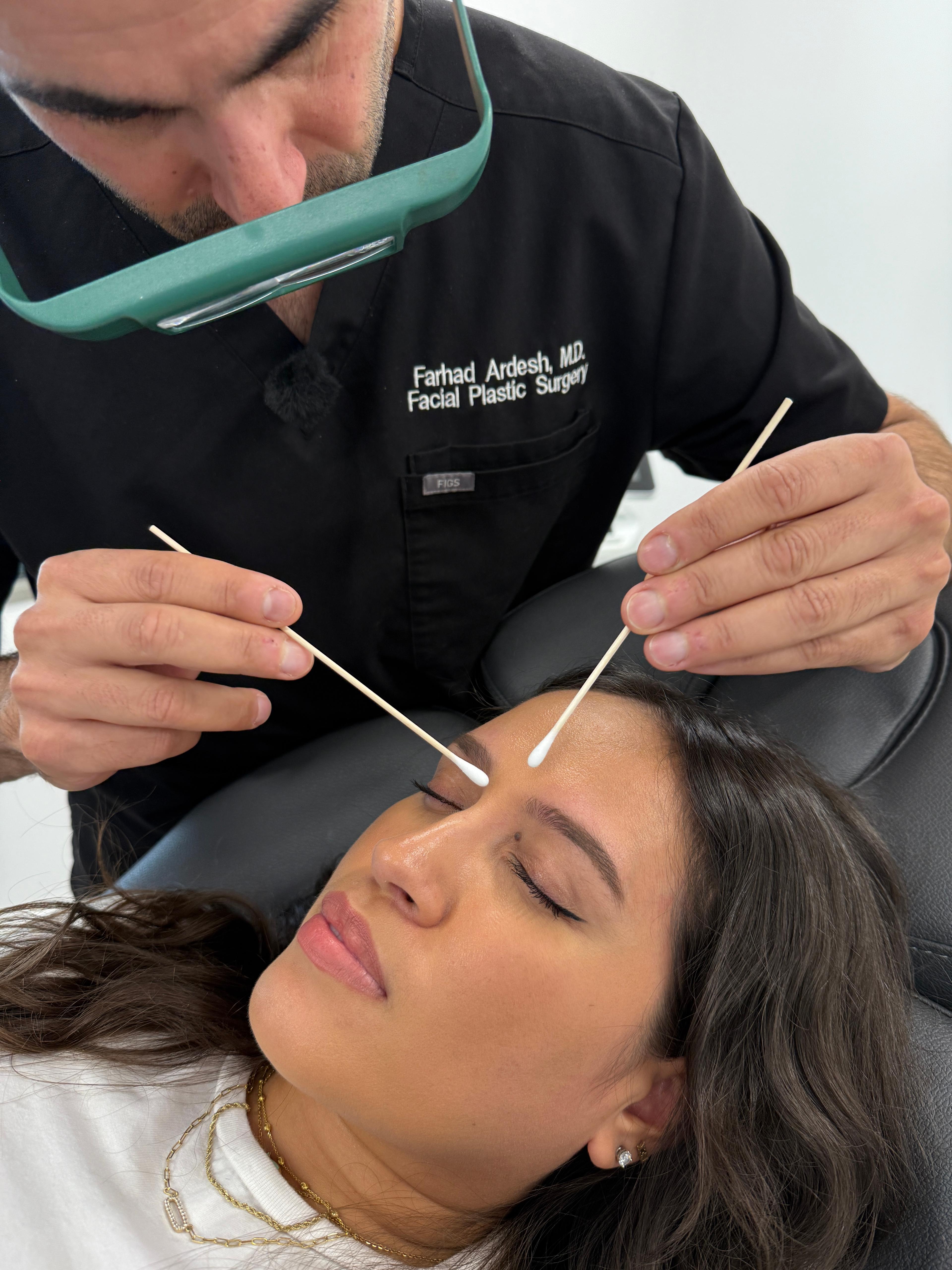
Dr. Ardesh examined the mole's size, shape and position, then evaluated the eyelid with the eyes open and closed. These movement checks help determine how the excision and closure can sit within the natural eyelid architecture.

Why this matters
The upper eyelid is thin, expressive and constantly moving. Evaluating the eye in both open and closed positions helps guide scar direction and closure planning around the natural crease.
03|Why This Area Is Different
The medial canthus is the inner corner of the eyelid beside the nose. It is a small, expressive area where scar direction and tissue behavior matter. A poorly oriented repair can create visible webbing, bunching or distortion.
Dr. Ardesh's experience with cosmetic upper blepharoplasty informed the planning and stitching strategy for this case, with attention to preserving a smooth contour and allowing the scar to sit naturally within the surrounding folds.
Planning Schematic
The incision and closure are planned in relation to the eyelid's natural folds and the inner corner of the eye.
Surgical Planning Priorities
Plan the repair in relation to eyelid folds so the scar can settle more discreetly.
Avoid a scar band that could pull across the inner corner of the eye.
Minimize dog ears, bunching and unnecessary scar length in a highly visible area.
Why this matters: In a thin, highly mobile area such as the upper eyelid, incision direction and tissue handling influence not only scar visibility, but also how naturally the eyelid moves and contours after healing.
04 | The Procedure
Dr. Ardesh performed Molectomy™ to completely remove the lesion while planning the repair around the delicate anatomy and movement of the upper eyelid. The goal was complete excision without sacrificing natural eyelid contour or creating unnecessary scar length.
1
Assess
Evaluate lesion position and eyelid movement with the eyes open and closed.
2
Plan
Align the repair with natural eyelid architecture and the medial canthus.
3
Remove
Perform Molectomy™ with complete excision of the lesion.
4
Close
Complete a refined repair designed to minimize scar length and bunching.
5
Review
Assess eyelid contour, movement and scar maturation at four months.
05 | Closure & Scar Planning
Special attention was given to minimizing the final scar length while avoiding dog ears, extra bunching of skin or distortion of the nearby eyelid contour. In this area, small irregularities can be more noticeable because the eyelid is thin, expressive and constantly moving.
Avoid
Plan for
Pathology confirmation
The removed tissue was sent for microscopic examination by a board-certified pathologist. Final pathology confirmed a benign melanocytic nevus, commonly known as a benign or noncancerous mole.
06 | Four-Month Outcome
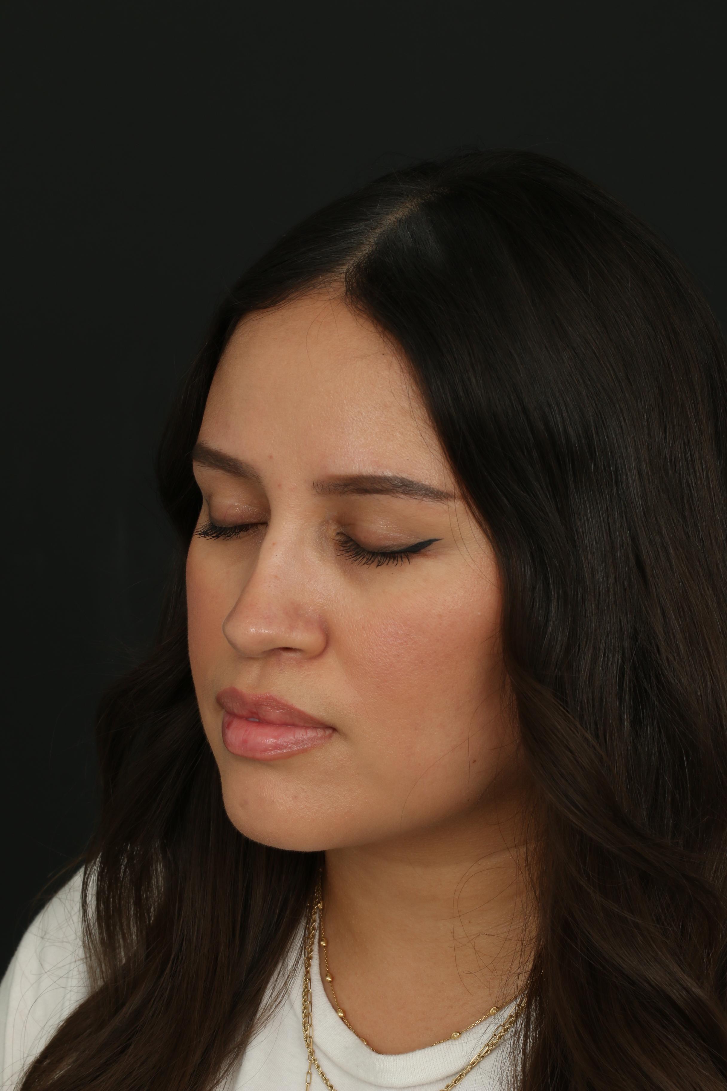
At the four-month follow-up, the raised, pigmented mole was no longer present. The upper eyelid shows a smooth, natural-looking contour, and the healing surgical scar blends well with the surrounding skin.

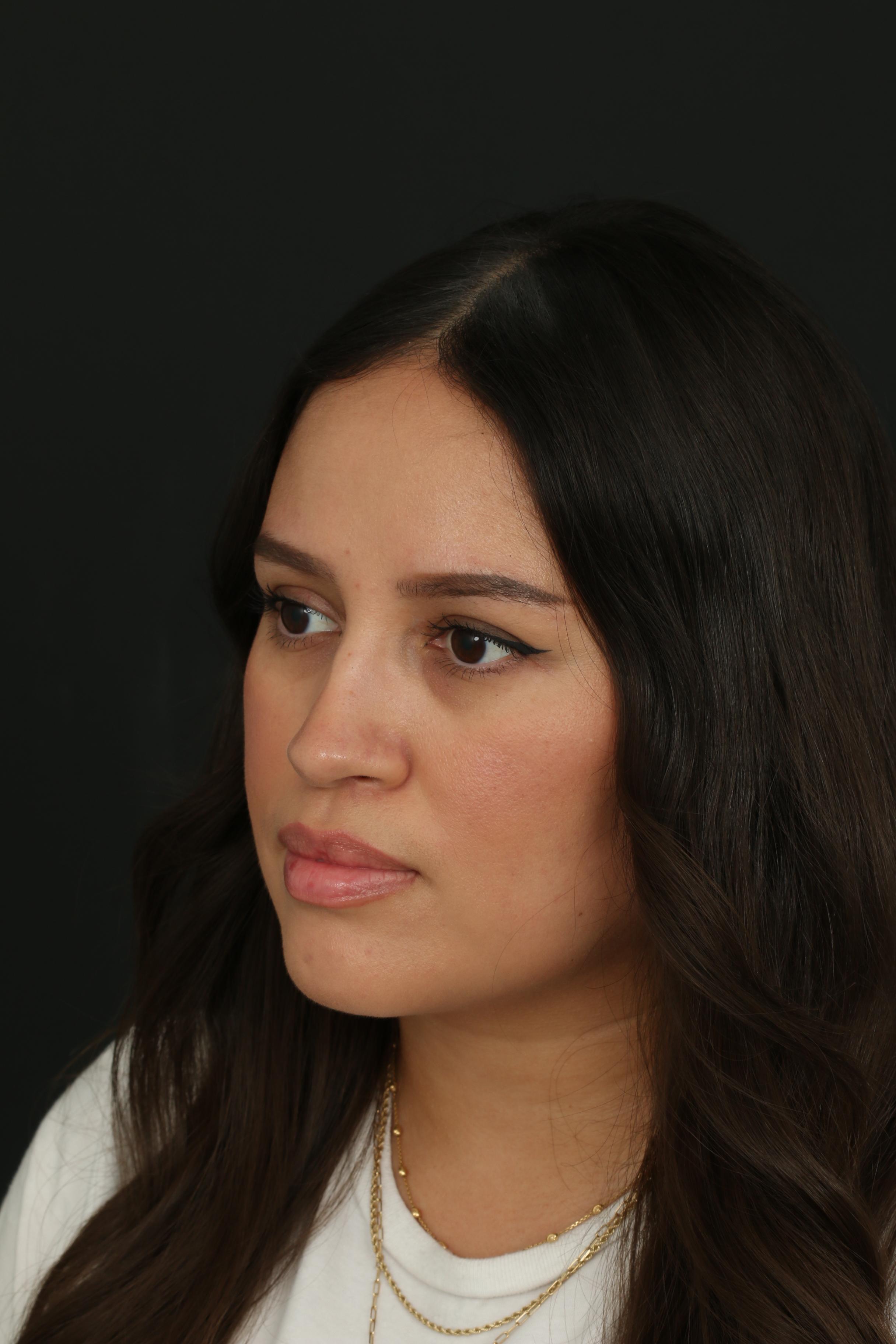
Four months after upper eyelid Molectomy™ — healed treatment area with preserved eyelid contour.
07 | Before & After
Matched views make it easier to compare the treatment area without relying on different camera angles or expressions.
08 | Video & Social Proof
These follow-up clips can add patient-facing and clinical context to the four-month result. Confirm the exact speaker and content of each source clip before publishing the final labels.
4-month follow-up
Use as the patient-facing follow-up module if the source clip contains her experience and result.
Patient perspective
Use the original clip if it contains the patient discussing treatment, recovery or satisfaction with the result.
Additional clinical video
Verify the exact speaker/content of this clip before assigning a more specific public-facing label.
Transformation summary
Before
Raised pigmented mole near the upper eyelid / medial canthus
Planning
Natural crease alignment, eyelid movement and webbing prevention
Procedure
Molectomy™ with refined eyelid-specific repair
Pathology
Benign melanocytic nevus
Follow-up
4 months
Outcome
Smooth natural-looking contour with a healing scar blending into surrounding skin
In this case, complete removal was only one part of the treatment plan. The lesion's location beside the medial canthus meant that eyelid movement, natural folds, scar direction and tissue behavior all needed to be considered before surgery.
The objective was a refined repair that respected the natural upper eyelid contour while minimizing webbing, bunching and unnecessary scar length.
Every mole and every treatment area is different. A consultation with Dr. Ardesh can help determine an appropriate treatment approach based on the lesion, surrounding anatomy and individual treatment goals.