
1
Assess
Evaluate movement, facial angles and the upper lip contour.
Mole Removal • Transformation Story
An eight-month transformation focused on complete removal, movement-based planning, multilayer closure and a natural-looking upper lip result.
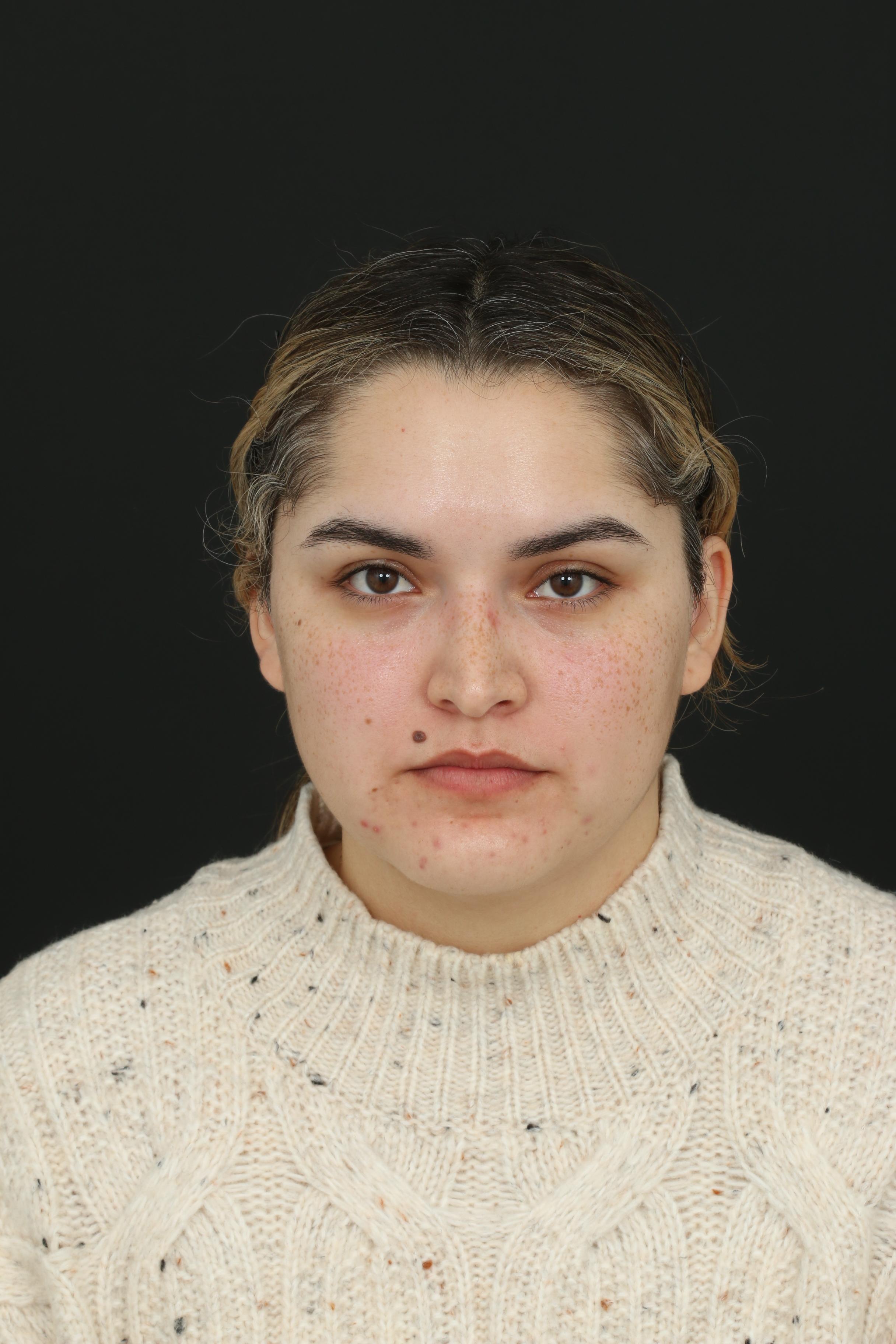
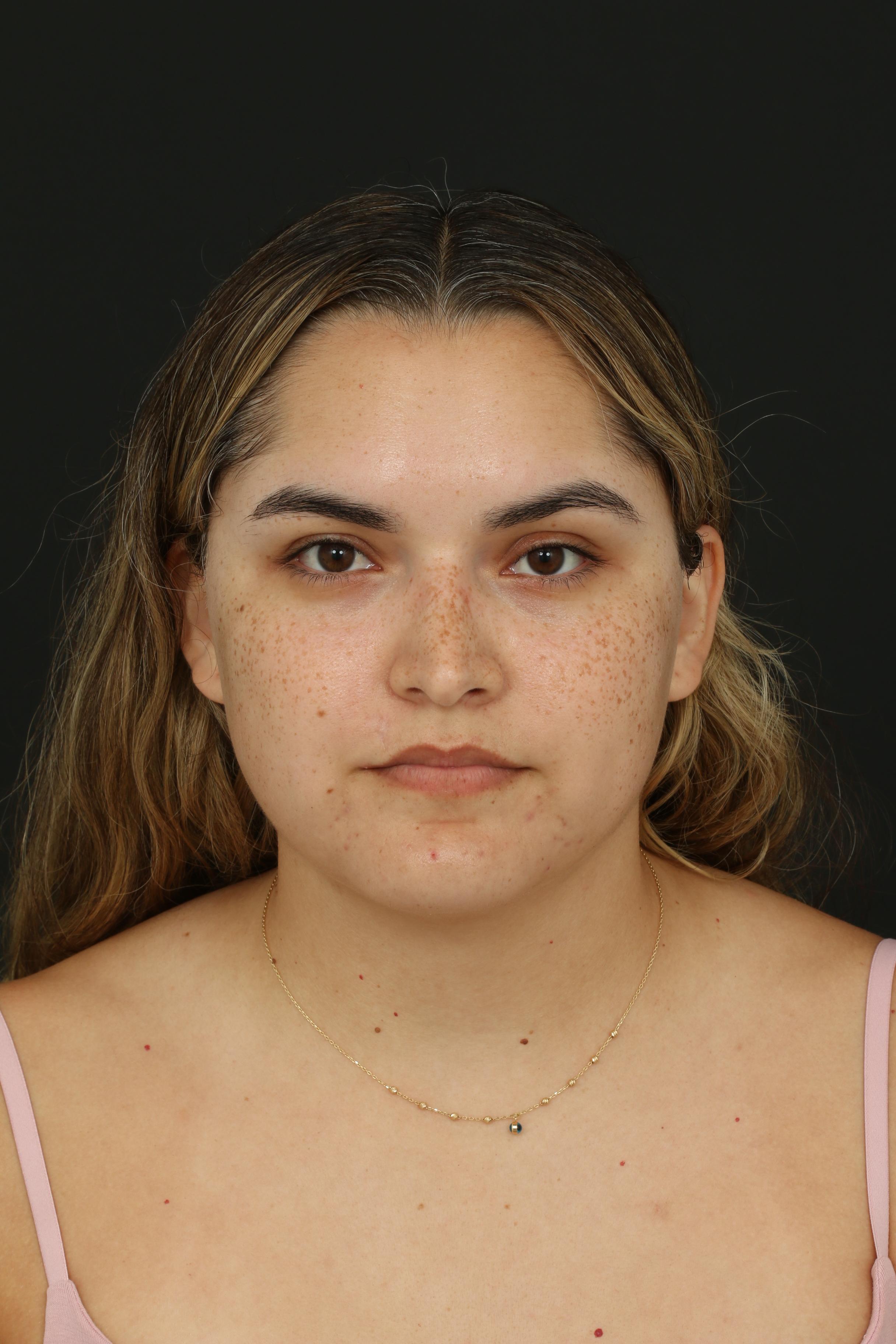
Actual patient shown before treatment and at eight-month follow-up. Individual healing and results vary.
Treatment
Molectomy™
Area
Upper lip
Follow-up
8 months
01 | Case at a Glance
This case follows an upper-lip Molectomy™ from multi-angle movement assessment through complete removal, multilayer closure, pathology and eight-month healing.
Primary Concern
Raised, dark-brown upper lip mole
Planning Focus
Normal facial movement from multiple angles, including smile, pucker and mouth opening
Closure
Multilayer closure with ultrafine sutures
Pathology
Benign nevus
Treatment
Molectomy™
Area Treated
Upper lip
Follow-up
8 months
Outcome
Smooth, natural-looking upper lip contour
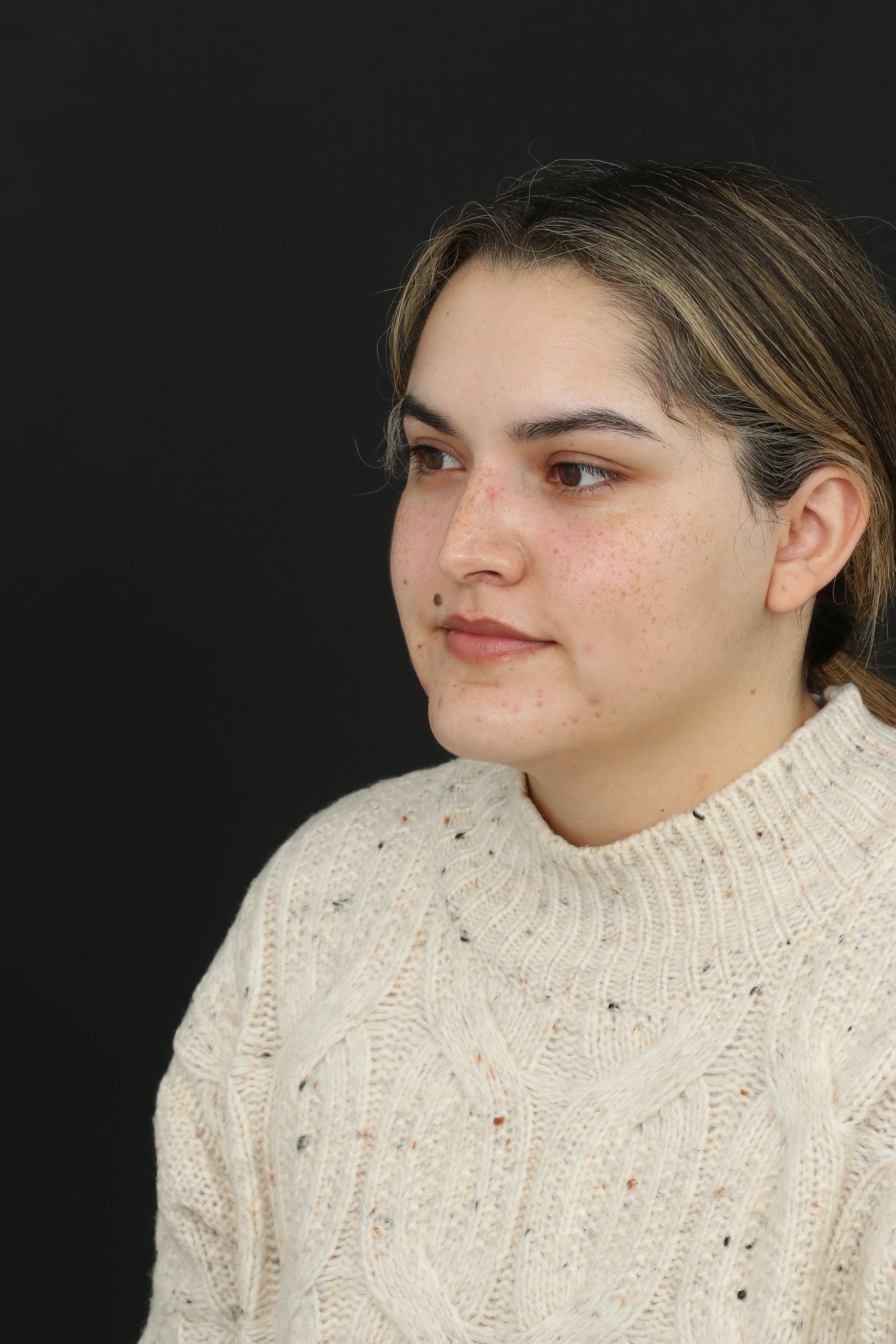
This 29-year-old woman wanted removal of a raised, dark-brown mole on her upper lip. Because the mole sat in a highly visible and expressive part of the face, planning needed to account for both complete removal and how the area would look in motion.
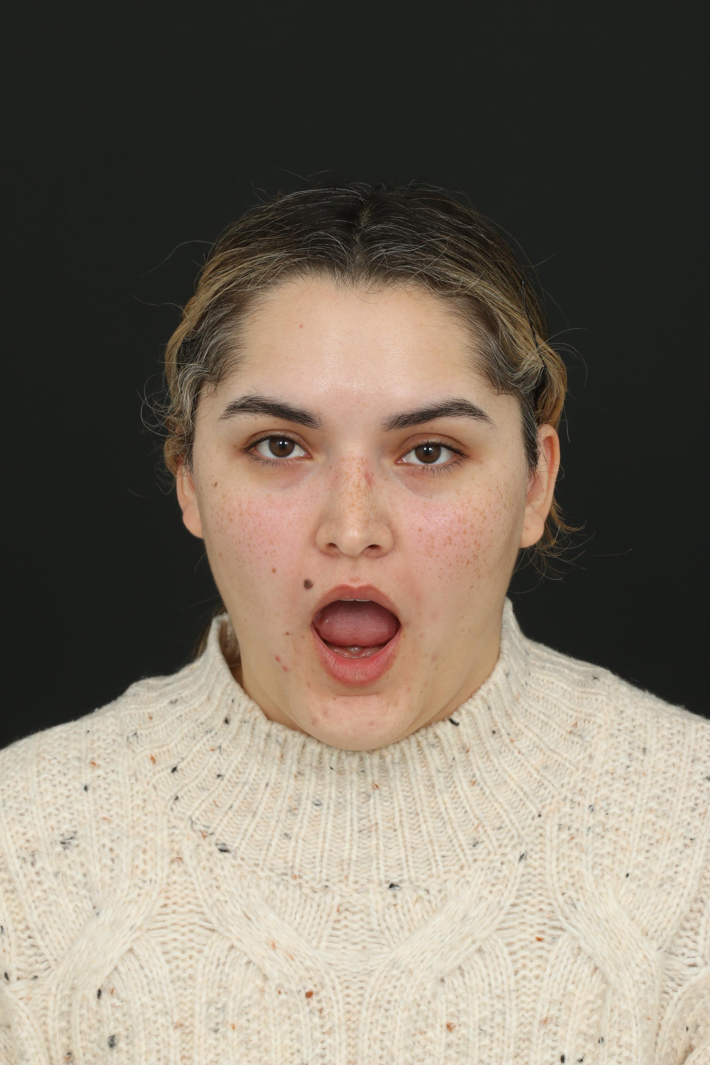
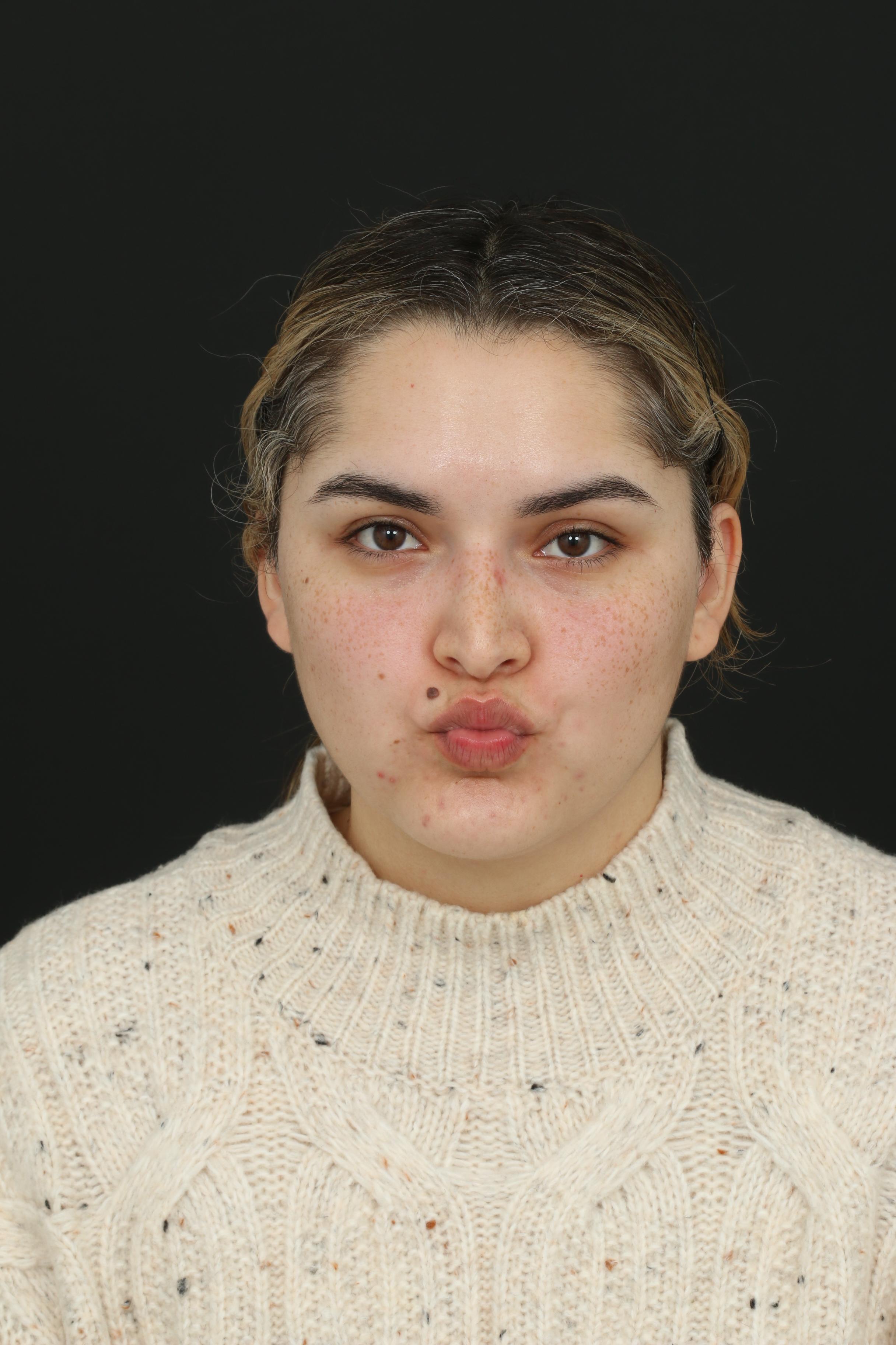
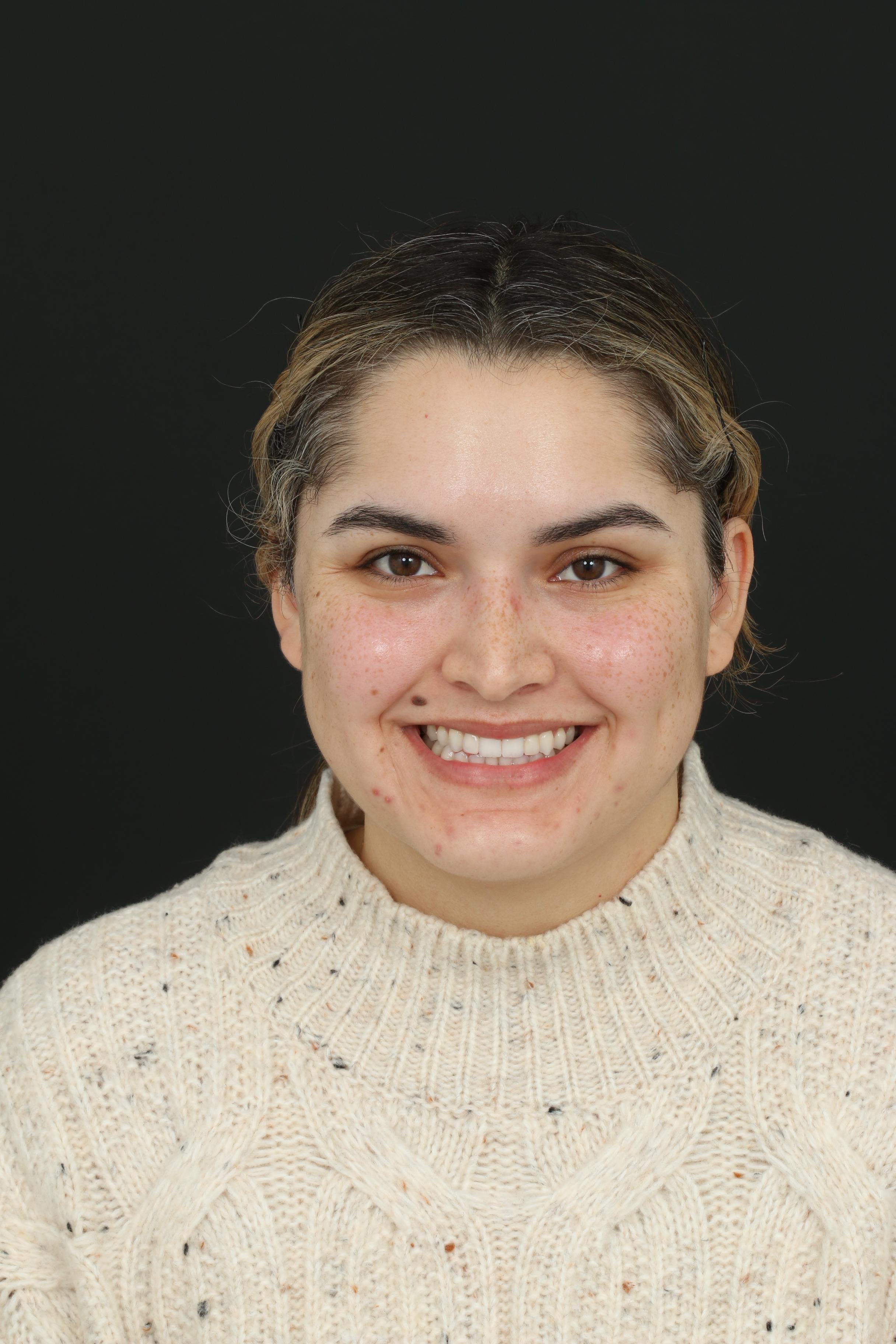
Smiling, puckering and opening the mouth all change the shape and tension of the upper lip, so the surgical design was assessed from multiple angles rather than from a single resting photograph.
02 | Assessment & Planning
Movement-based planning
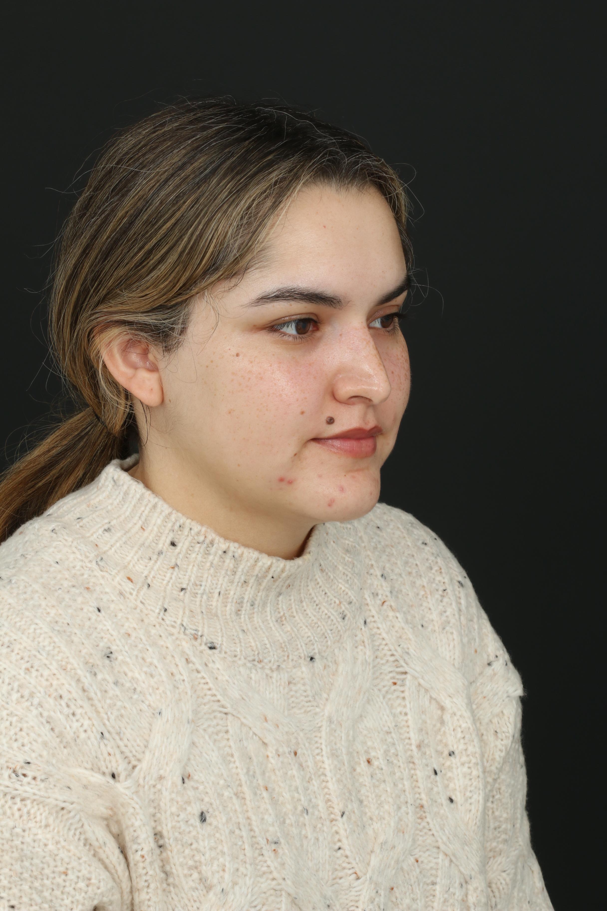
Dr. Ardesh evaluated the upper lip from multiple angles and during normal facial movement. Smiling, puckering and opening the mouth change the shape and tension of the lip, so these expressions were considered when planning the excision and surgical repair.
03 | Surgical Design
The goal was to completely remove the mole while planning the final repair so the scar could remain as inconspicuous as possible across different expressions and viewing angles.
The lesion position, surrounding skin, natural lip shape and direction of movement were considered together before treatment rather than treating the mole as an isolated surface feature.
04 | The Molectomy™ Approach
Dr. Ardesh performed his signature Molectomy™ to completely remove the mole, including its deeper components and unwanted hair follicles. The procedure was planned with the long-term aesthetic result in mind rather than focusing only on the visible surface.
1
Assess
Evaluate movement, facial angles and the upper lip contour.
2
Remove
Completely excise the mole, including deeper components and unwanted hair follicles.
3
Close
Use a multilayer closure with ultrafine sutures to distribute tension through deeper tissues.
4
Heal
Allow the scar and upper lip contour to mature through the eight-month follow-up.
Closure strategy
Following removal, a multilayer closure with ultrafine sutures was performed to support healing and help minimize the final scar. This layered approach distributes tension through deeper tissues rather than relying only on the skin surface.
Tension Control
Support the repair during normal facial movement.
Short, Refined Scar
Avoid unnecessary scar length while respecting the anatomy.
Natural Contour
Preserve the upper lip shape from multiple viewing angles.
05 | Clinical Confirmation
The removed mole was sent to a laboratory for microscopic evaluation by a board-certified pathologist. The tissue was confirmed to be a benign nevus.
Molectomy™ includes not only removal and reconstruction, but also appropriate evaluation of the excised tissue when indicated. This provides a documented microscopic diagnosis after the procedure.
06 | 8-Month Follow-Up
At eight months after surgery, the raised mole is no longer present and the upper lip maintains a smooth, natural-looking contour. The result can be assessed both at rest and during expression, reflecting the importance of movement-based planning from the beginning.
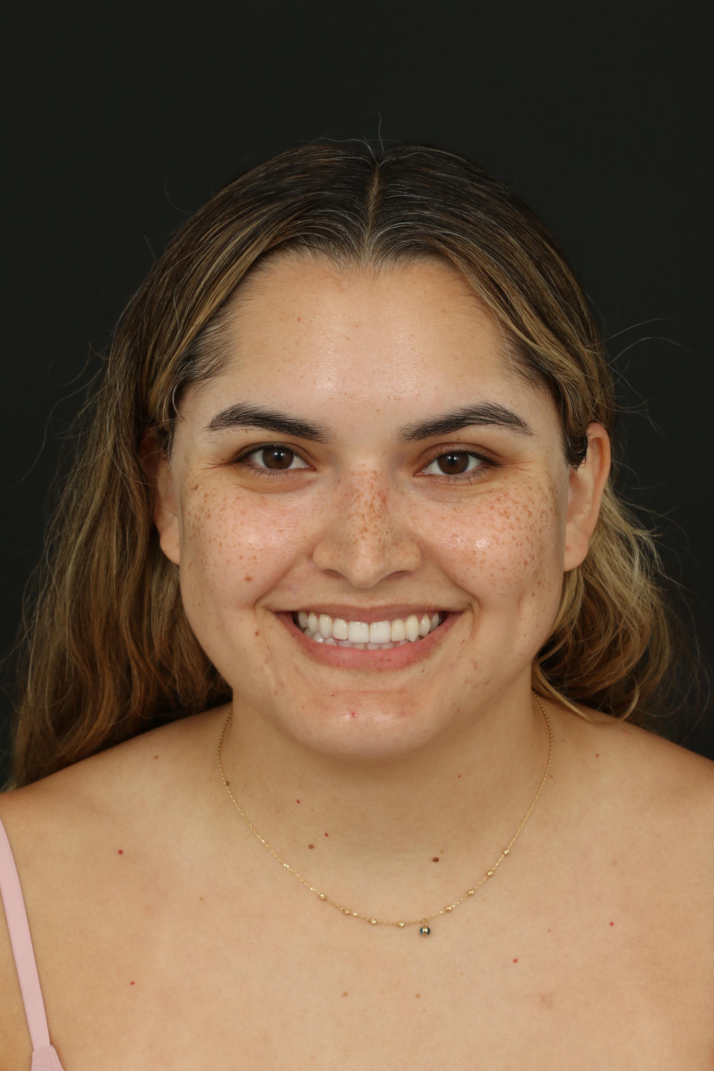
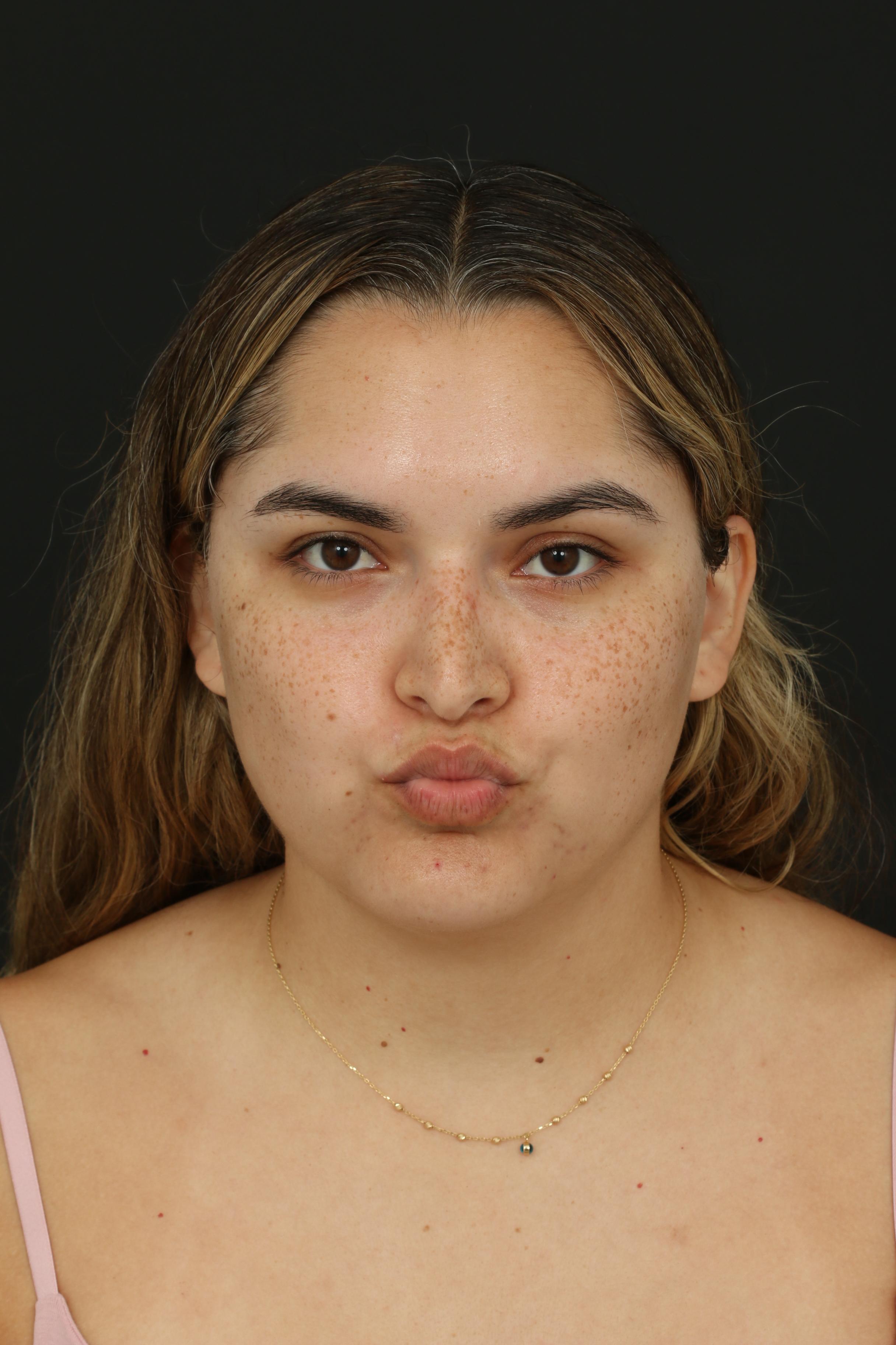
Eight months after upper lip Molectomy™ — result shown at rest and during expression.
07 | Before & After
Matching facial expressions help demonstrate the result in motion rather than relying on a single static photograph.
08 | Video & Social Proof
The physician interview and patient testimonial close the clinical story with both the treatment rationale and the patient's own experience at eight months.
Doctor perspective
The physician perspective on movement-based planning, complete removal, pathology, multilayer closure and the eight-month result.
Patient testimonial
The patient shares her experience and perspective on the long-term result.
Supporting doctor + patient footage
Transformation summary
Before
Raised, dark-brown upper lip mole
Planning
Smile, pucker, open-mouth and multi-angle assessment
Procedure
Molectomy™ with complete removal
Closure
Multilayer repair with ultrafine sutures
Pathology
Benign nevus
Follow-up
8 months
Outcome
Smooth, natural-looking upper lip contour
The upper lip changes shape with smiling, puckering, speaking and mouth opening. In this case, the treatment plan therefore extended beyond removing the visible mole and included planning the scar and closure around those normal movements.
The objective was complete removal with a layered repair that supported healing while preserving a natural upper lip contour at rest and during expression.
Every mole and every treatment area is different. A consultation with Dr. Ardesh can help determine the most appropriate approach based on the characteristics of the mole, its location, facial movement and individual treatment goals.